Example of JACC: Cardiovascular Imaging format
Recent searches

Example of JACC: Cardiovascular Imaging format Example of JACC: Cardiovascular Imaging format Example of JACC: Cardiovascular Imaging format Example of JACC: Cardiovascular Imaging format Example of JACC: Cardiovascular Imaging format Example of JACC: Cardiovascular Imaging format Example of JACC: Cardiovascular Imaging format Example of JACC: Cardiovascular Imaging format Example of JACC: Cardiovascular Imaging format Example of JACC: Cardiovascular Imaging format Example of JACC: Cardiovascular Imaging format Example of JACC: Cardiovascular Imaging format Example of JACC: Cardiovascular Imaging format Example of JACC: Cardiovascular Imaging format Example of JACC: Cardiovascular Imaging format Example of JACC: Cardiovascular Imaging format Example of JACC: Cardiovascular Imaging format
Sample paper formatted on SciSpace - SciSpace
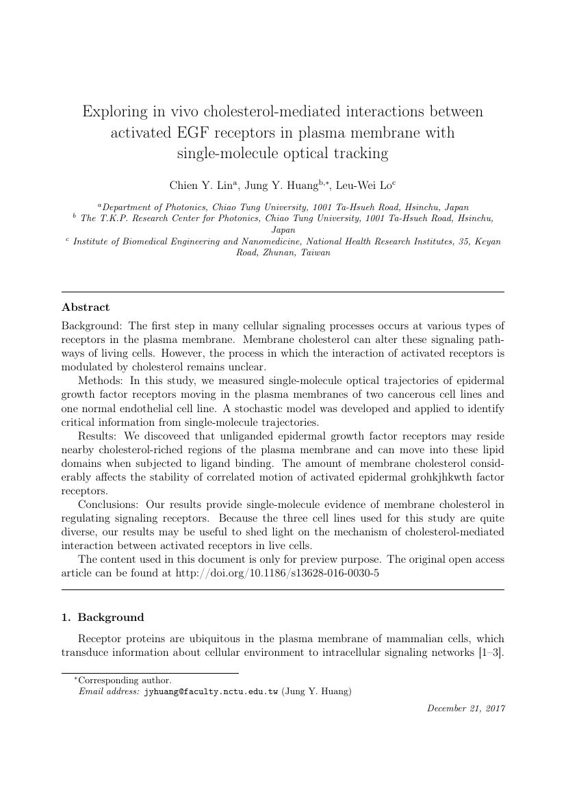
This content is only for preview purposes. The original open access content can be found here.
Look Inside
Example of JACC: Cardiovascular Imaging format Example of JACC: Cardiovascular Imaging format Example of JACC: Cardiovascular Imaging format Example of JACC: Cardiovascular Imaging format Example of JACC: Cardiovascular Imaging format Example of JACC: Cardiovascular Imaging format Example of JACC: Cardiovascular Imaging format Example of JACC: Cardiovascular Imaging format Example of JACC: Cardiovascular Imaging format Example of JACC: Cardiovascular Imaging format Example of JACC: Cardiovascular Imaging format Example of JACC: Cardiovascular Imaging format Example of JACC: Cardiovascular Imaging format Example of JACC: Cardiovascular Imaging format Example of JACC: Cardiovascular Imaging format Example of JACC: Cardiovascular Imaging format Example of JACC: Cardiovascular Imaging format
Sample paper formatted on SciSpace - SciSpace
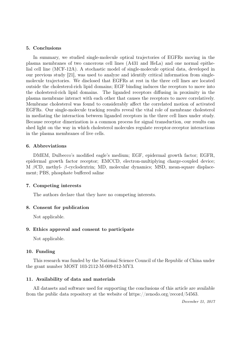
This content is only for preview purposes. The original open access content can be found here.
open access Open Access
recommended Recommended

JACC: Cardiovascular Imaging — Template for authors

Publisher: Elsevier
Categories Rank Trend in last 3 yrs
Radiology, Nuclear Medicine and Imaging #4 of 288 down down by 2 ranks
Cardiology and Cardiovascular Medicine #9 of 317 down down by 2 ranks
journal-quality-icon Journal quality:
High
calendar-icon Last 4 years overview: 594 Published Papers | 9953 Citations
indexed-in-icon Indexed in: Scopus
last-updated-icon Last updated: 18/07/2020
Related journals
Insights
General info
Top papers
Popular templates
Get started guide
Why choose from SciSpace
FAQ

Related Journals

open access Open Access
recommended Recommended

SAGE

Quality:  
High
CiteRatio: 5.5
SJR: 1.72
SNIP: 1.405
open access Open Access
recommended Recommended

Springer

Quality:  
High
CiteRatio: 9.0
SJR: 2.558
SNIP: 2.194
open access Open Access

Elsevier

Quality:  
High
CiteRatio: 4.8
SJR: 1.382
SNIP: 1.312
open access Open Access
recommended Recommended

Elsevier

Quality:  
High
CiteRatio: 8.3
SJR: 2.95
SNIP: 2.747

Journal Performance & Insights

Impact Factor

CiteRatio

Determines the importance of a journal by taking a measure of frequency with which the average article in a journal has been cited in a particular year.

A measure of average citations received per peer-reviewed paper published in the journal.

12.74

16% from 2018

Impact factor for JACC: Cardiovascular Imaging from 2016 - 2019
Year Value
2019 12.74
2018 10.975
2017 10.247
2016 10.189
graph view Graph view
table view Table view

16.8

14% from 2019

CiteRatio for JACC: Cardiovascular Imaging from 2016 - 2020
Year Value
2020 16.8
2019 14.8
2018 14.4
2017 14.0
2016 13.6
graph view Graph view
table view Table view

insights Insights

  • Impact factor of this journal has increased by 16% in last year.
  • This journal’s impact factor is in the top 10 percentile category.

insights Insights

  • CiteRatio of this journal has increased by 14% in last years.
  • This journal’s CiteRatio is in the top 10 percentile category.

SCImago Journal Rank (SJR)

Source Normalized Impact per Paper (SNIP)

Measures weighted citations received by the journal. Citation weighting depends on the categories and prestige of the citing journal.

Measures actual citations received relative to citations expected for the journal's category.

5.79

18% from 2019

SJR for JACC: Cardiovascular Imaging from 2016 - 2020
Year Value
2020 5.79
2019 4.902
2018 5.102
2017 4.66
2016 5.038
graph view Graph view
table view Table view

3.409

13% from 2019

SNIP for JACC: Cardiovascular Imaging from 2016 - 2020
Year Value
2020 3.409
2019 3.029
2018 2.723
2017 2.77
2016 2.741
graph view Graph view
table view Table view

insights Insights

  • SJR of this journal has increased by 18% in last years.
  • This journal’s SJR is in the top 10 percentile category.

insights Insights

  • SNIP of this journal has increased by 13% in last years.
  • This journal’s SNIP is in the top 10 percentile category.
JACC: Cardiovascular Imaging

Guideline source: View

All company, product and service names used in this website are for identification purposes only. All product names, trademarks and registered trademarks are property of their respective owners.

Use of these names, trademarks and brands does not imply endorsement or affiliation. Disclaimer Notice

Elsevier

JACC: Cardiovascular Imaging

JACC: Cardiovascular Imaging provides readers with a broad, balanced view of all aspects of cardiovascular imaging. The Journal includes original clinical research on non-invasive and invasive imaging techniques including echocardiography, CT, CMR, nuclear, optical imaging, an...... Read More

Medicine

i
Last updated on
18 Jul 2020
i
ISSN
1936-878X
i
Impact Factor
High - 2.101
i
Acceptance Rate
15%
i
Open Access
No
i
Sherpa RoMEO Archiving Policy
Green faq
i
Plagiarism Check
Available via Turnitin
i
Endnote Style
Download Available
i
Bibliography Name
elsarticle-num
i
Citation Type
Numbered
[25]
i
Bibliography Example
G. E. Blonder, M. Tinkham, T. M. Klapwijk, Transition from metallic to tunneling regimes in superconducting microconstrictions: Excess current, charge imbalance, and supercurrent conversion, Phys. Rev. B 25 (7) (1982) 4515–4532. URL 10.1103/PhysRevB.25.4515

Top papers written in this journal

open accessOpen access Journal Article DOI: 10.1016/J.JCMG.2010.10.008
Left ventricular remodeling in heart failure: current concepts in clinical significance and assessment.
Marvin A. Konstam1, Daniel G. Kramer1, Ayan R. Patel1, Martin S. Maron1, James E. Udelson1

Abstract:

Ventricular remodeling, first described in animal models of left ventricular (LV) stress and injury, occurs progressively in untreated patients after large myocardial infarction and in those with dilated forms of cardiomyopathy. The gross pathologic changes of increased LV volume and perturbation in the normal elliptical LV c... Ventricular remodeling, first described in animal models of left ventricular (LV) stress and injury, occurs progressively in untreated patients after large myocardial infarction and in those with dilated forms of cardiomyopathy. The gross pathologic changes of increased LV volume and perturbation in the normal elliptical LV chamber configuration is driven, on a histologic level, by myocyte hypertrophy and apoptosis and by increased interstitial collagen. Each of the techniques used for tracking this process—echocardiography, radionuclide ventriculography, and cardiac magnetic resonance—carries advantages and disadvantages. Numerous investigations have demonstrated the value of LV volume measurement at a single time-point and over time in predicting clinical outcomes in patients with heart failure and in those after myocardial infarction. The structural pattern of LV remodeling and evidence of scarring on cardiac magnetic resonance have additional prognostic value. Beyond the impact of abnormal cardiac structure on cardiovascular events, the relationship between LV remodeling and clinical outcomes is likely linked through common local and systemic factors driving vascular as well as myocardial pathology. As demonstrated by a recent meta-analysis of heart failure trials, LV volume stands out among surrogate markers as strongly correlating with the impact of a particular drug or device therapy on patient survival. These findings substantiate the importance of ventricular remodeling as central in the pathophysiology of advancing heart failure and support the role of measures of LV remodeling in the clinical investigation of novel heart failure treatments. read more read less

Topics:

Ventricular remodeling (67%)67% related to the paper, Heart failure (59%)59% related to the paper, Cardiomyopathy (58%)58% related to the paper, Myocardial infarction (57%)57% related to the paper, Radionuclide ventriculography (57%)57% related to the paper
605 Citations
open accessOpen access Journal Article DOI: 10.1016/J.JCMG.2008.12.031
Diagnostic and Prognostic Value of Absence of Coronary Artery Calcification

Abstract:

Objectives In this study, we systematically assessed the diagnostic and prognostic value of absence of coronary artery calcification (CAC) in asymptomatic and symptomatic individuals Background Presence of CAC is a well-established marker of coronary plaque burden and is associated with a higher risk of adverse cardiovascular... Objectives In this study, we systematically assessed the diagnostic and prognostic value of absence of coronary artery calcification (CAC) in asymptomatic and symptomatic individuals Background Presence of CAC is a well-established marker of coronary plaque burden and is associated with a higher risk of adverse cardiovascular outcomes Absence of CAC has been suggested to be associated with a very low risk of significant coronary artery disease, as well as minimal risk of future events Methods We searched online databases (eg, PubMed and MEDLINE) for original research articles published in English between January 1990 and March 2008 examining the diagnostic and prognostic utility of CAC Results A systematic review of published articles revealed 49 studies that fulfilled our criteria for inclusion These included 13 studies assessing the relationship of CAC with adverse cardiovascular outcomes in 64,873 asymptomatic patients In this cohort, 146 of 25,903 patients without CAC (056%) had a cardiovascular event during a mean follow-up period of 51 months In the 7 studies assessing the prognostic value of CAC in a symptomatic population, 180% of patients without CAC had a cardiovascular event Overall, 18 studies demonstrated that the presence of any CAC had a pooled sensitivity and negative predictive value of 98% and 93%, respectively, for detection of significant coronary artery disease on invasive coronary angiography In 4,870 individuals undergoing myocardial perfusion and CAC testing, in the absence of CAC, only 6% demonstrated any sign of ischemia Finally, 3 studies demonstrated that absence of CAC had a negative predictive value of 99% for ruling out acute coronary syndrome Conclusions On the basis of our review of more than 85,000 patients, we conclude that the absence of CAC is associated with a very low risk of future cardiovascular events, with modest incremental value of other diagnostic tests in this very low-risk group read more read less

Topics:

Coronary artery disease (56%)56% related to the paper, Acute coronary syndrome (53%)53% related to the paper, Predictive value of tests (52%)52% related to the paper, Population (50%)50% related to the paper
602 Citations
open accessOpen access Journal Article DOI: 10.1016/J.JCMG.2010.11.015
Evaluation of techniques for the quantification of myocardial scar of differing etiology using cardiac magnetic resonance

Abstract:

Objectives The aim of this study was to compare the reproducibility of 7 late gadolinium enhancement (LGE) quantification techniques across 3 conditions in which LGE is known to be important: acute myocardial infarction (AMI), chronic myocardial infarction (CMI), and hypertrophic cardiomyopathy (HCM) Background LGE by cardiac... Objectives The aim of this study was to compare the reproducibility of 7 late gadolinium enhancement (LGE) quantification techniques across 3 conditions in which LGE is known to be important: acute myocardial infarction (AMI), chronic myocardial infarction (CMI), and hypertrophic cardiomyopathy (HCM) Background LGE by cardiac magnetic resonance is the gold-standard technique for assessing myocardial scar No consensus exists on the best method for its quantification, and research in this area is scant Techniques include manual quantification, thresholding by 2, 3, 4, 5, or 6 SDs above remote myocardium, and the full width at half maximum (FWHM) technique To date, LGE has been linked to outcome in 3 conditions: AMI, CMI, and HCM Methods Sixty patients with 3 LGE etiologies (AMI, n = 20; CMI, n = 20; HCM, n = 20) were scanned for LGE LGE volume was quantified using the 7 techniques Mean LGE volume, interobserver and intraobserver reproducibility, and impact on sample size were assessed Results LGE volume varied significantly with the quantification method used There was no statistically significant difference between LGE volume by the FWHM, manual, and 6-SD or 5-SD techniques The 2-SD technique generated LGE volumes up to 2 times higher than the FWHM, 6-SD, and manual techniques The reproducibility of all techniques was worse in HCM than AMI or CMI The FWHM technique was the most reproducible in all 3 conditions compared with any other method (p Conclusions Regardless of the disease under study, the FWHM technique for LGE quantification gives LGE volume mean results similar to manual quantification and is statistically the most reproducible, reducing required sample sizes by up to one-half read more read less
528 Citations
open accessOpen access Journal Article DOI: 10.1016/J.JCMG.2008.02.006
Twist Mechanics of the Left Ventricle: Principles and Application
Partho P. Sengupta1, A. Jamil Tajik1, Krishnaswamy Chandrasekaran1, Bijoy K. Khandheria1

Abstract:

Left ventricular (LV) twist or torsion represents the mean longitudinal gradient of the net difference in clockwise and counterclockwise rotation of the LV apex and base, as viewed from LV apex. Twist during ejection predominantly deforms the subendocardial fiber matrix, resulting in storage of potential energy. Subsequent re... Left ventricular (LV) twist or torsion represents the mean longitudinal gradient of the net difference in clockwise and counterclockwise rotation of the LV apex and base, as viewed from LV apex. Twist during ejection predominantly deforms the subendocardial fiber matrix, resulting in storage of potential energy. Subsequent recoil of twist deformation is associated with the release of restoring forces, which contributes to LV diastolic relaxation and early diastolic filling. Noninvasive techniques such as magnetic resonance imaging and echocardiography are useful for understanding LV twist dynamics in clinical settings, and data regarding their relative merits and pitfalls are rapidly accumulating. This review is a focused update on the current and evolving applications of LV twist mechanics in clinical cardiology. First, the theoretical framework for understanding the physiological sequence of LV twist during a cardiac cycle is presented. Second, variations in LV twist encountered in different experimental and clinical situations are discussed. Finally, the review presents an algorithm for routine application of LV twist in clinical differentiation of patterns of LV dysfunction encountered in day-to-day practice. read more read less
527 Citations
Journal Article DOI: 10.1016/J.JCMG.2012.11.013
Noncontrast T1 mapping for the diagnosis of cardiac amyloidosis.

Abstract:

Objectives This study sought to explore the potential role of noncontrast myocardial T1 mapping for detection of cardiac involvement in patients with primary amyloid light-chain (AL) amyloidosis. Background Cardiac involvement carries a poor prognosis in systemic AL amyloidosis. Late gadolinium enhancement (LGE) cardiac magne... Objectives This study sought to explore the potential role of noncontrast myocardial T1 mapping for detection of cardiac involvement in patients with primary amyloid light-chain (AL) amyloidosis. Background Cardiac involvement carries a poor prognosis in systemic AL amyloidosis. Late gadolinium enhancement (LGE) cardiac magnetic resonance (CMR) is useful for the detection of cardiac amyloid, but characteristic LGE patterns do not always occur or they appear late in the disease. Noncontrast characterization of amyloidotic myocardium with T1 mapping may improve disease detection. Furthermore, quantitative assessment of myocardial amyloid load would be of great value. Methods Fifty-three AL amyloidosis patients (14 with no cardiac involvement, 11 with possible involvement, and 28 with definite cardiac involvement based on standard biomarker and echocardiographic criteria) underwent CMR (1.5-T) including noncontrast T1 mapping (shortened modified look-locker inversion recovery [ShMOLLI] sequence) and LGE imaging. These were compared with 36 healthy volunteers and 17 patients with aortic stenosis and a comparable degree of left ventricular hypertrophy as the cardiac amyloid patients. Results Myocardial T1 was significantly elevated in cardiac AL amyloidosis patients (1,140 ± 61 ms) compared to normal subjects (958 ± 20 ms, p Conclusions Noncontrast T1 mapping has high diagnostic accuracy for detecting cardiac AL amyloidosis, correlates well with markers of systolic and diastolic dysfunction, and is potentially more sensitive for detecting early disease than LGE imaging. Elevated myocardial T1 may represent a direct marker of cardiac amyloid load. Further studies are needed to assess the prognostic significance of T1 elevation. read more read less

Topics:

Cardiac amyloidosis (69%)69% related to the paper, AL amyloidosis (60%)60% related to the paper, Amyloidosis (60%)60% related to the paper, Primary systemic amyloidosis (58%)58% related to the paper, Myocardial infarction (53%)53% related to the paper
525 Citations
Author Pic

SciSpace is a very innovative solution to the formatting problem and existing providers, such as Mendeley or Word did not really evolve in recent years.

- Andreas Frutiger, Researcher, ETH Zurich, Institute for Biomedical Engineering

Get MS-Word and LaTeX output to any Journal within seconds
1
Choose a template
Select a template from a library of 40,000+ templates
2
Import a MS-Word file or start fresh
It takes only few seconds to import
3
View and edit your final output
SciSpace will automatically format your output to meet journal guidelines
4
Submit directly or Download
Submit to journal directly or Download in PDF, MS Word or LaTeX

(Before submission check for plagiarism via Turnitin)

clock Less than 3 minutes

What to expect from SciSpace?

Speed and accuracy over MS Word

''

With SciSpace, you do not need a word template for JACC: Cardiovascular Imaging.

It automatically formats your research paper to Elsevier formatting guidelines and citation style.

You can download a submission ready research paper in pdf, LaTeX and docx formats.

Time comparison

Time taken to format a paper and Compliance with guidelines

Plagiarism Reports via Turnitin

SciSpace has partnered with Turnitin, the leading provider of Plagiarism Check software.

Using this service, researchers can compare submissions against more than 170 million scholarly articles, a database of 70+ billion current and archived web pages. How Turnitin Integration works?

Turnitin Stats
Publisher Logos

Freedom from formatting guidelines

One editor, 100K journal formats – world's largest collection of journal templates

With such a huge verified library, what you need is already there.

publisher-logos

Easy support from all your favorite tools

JACC: Cardiovascular Imaging format uses elsarticle-num citation style.

Automatically format and order your citations and bibliography in a click.

SciSpace allows imports from all reference managers like Mendeley, Zotero, Endnote, Google Scholar etc.

Frequently asked questions

1. Can I write JACC: Cardiovascular Imaging in LaTeX?

Absolutely not! Our tool has been designed to help you focus on writing. You can write your entire paper as per the JACC: Cardiovascular Imaging guidelines and auto format it.

2. Do you follow the JACC: Cardiovascular Imaging guidelines?

Yes, the template is compliant with the JACC: Cardiovascular Imaging guidelines. Our experts at SciSpace ensure that. If there are any changes to the journal's guidelines, we'll change our algorithm accordingly.

3. Can I cite my article in multiple styles in JACC: Cardiovascular Imaging?

Of course! We support all the top citation styles, such as APA style, MLA style, Vancouver style, Harvard style, and Chicago style. For example, when you write your paper and hit autoformat, our system will automatically update your article as per the JACC: Cardiovascular Imaging citation style.

4. Can I use the JACC: Cardiovascular Imaging templates for free?

Sign up for our free trial, and you'll be able to use all our features for seven days. You'll see how helpful they are and how inexpensive they are compared to other options, Especially for JACC: Cardiovascular Imaging.

5. Can I use a manuscript in JACC: Cardiovascular Imaging that I have written in MS Word?

Yes. You can choose the right template, copy-paste the contents from the word document, and click on auto-format. Once you're done, you'll have a publish-ready paper JACC: Cardiovascular Imaging that you can download at the end.

6. How long does it usually take you to format my papers in JACC: Cardiovascular Imaging?

It only takes a matter of seconds to edit your manuscript. Besides that, our intuitive editor saves you from writing and formatting it in JACC: Cardiovascular Imaging.

7. Where can I find the template for the JACC: Cardiovascular Imaging?

It is possible to find the Word template for any journal on Google. However, why use a template when you can write your entire manuscript on SciSpace , auto format it as per JACC: Cardiovascular Imaging's guidelines and download the same in Word, PDF and LaTeX formats? Give us a try!.

8. Can I reformat my paper to fit the JACC: Cardiovascular Imaging's guidelines?

Of course! You can do this using our intuitive editor. It's very easy. If you need help, our support team is always ready to assist you.

9. JACC: Cardiovascular Imaging an online tool or is there a desktop version?

SciSpace's JACC: Cardiovascular Imaging is currently available as an online tool. We're developing a desktop version, too. You can request (or upvote) any features that you think would be helpful for you and other researchers in the "feature request" section of your account once you've signed up with us.

10. I cannot find my template in your gallery. Can you create it for me like JACC: Cardiovascular Imaging?

Sure. You can request any template and we'll have it setup within a few days. You can find the request box in Journal Gallery on the right side bar under the heading, "Couldn't find the format you were looking for like JACC: Cardiovascular Imaging?”

11. What is the output that I would get after using JACC: Cardiovascular Imaging?

After writing your paper autoformatting in JACC: Cardiovascular Imaging, you can download it in multiple formats, viz., PDF, Docx, and LaTeX.

12. Is JACC: Cardiovascular Imaging's impact factor high enough that I should try publishing my article there?

To be honest, the answer is no. The impact factor is one of the many elements that determine the quality of a journal. Few of these factors include review board, rejection rates, frequency of inclusion in indexes, and Eigenfactor. You need to assess all these factors before you make your final call.

13. What is Sherpa RoMEO Archiving Policy for JACC: Cardiovascular Imaging?

SHERPA/RoMEO Database

We extracted this data from Sherpa Romeo to help researchers understand the access level of this journal in accordance with the Sherpa Romeo Archiving Policy for JACC: Cardiovascular Imaging. The table below indicates the level of access a journal has as per Sherpa Romeo's archiving policy.

RoMEO Colour Archiving policy
Green Can archive pre-print and post-print or publisher's version/PDF
Blue Can archive post-print (ie final draft post-refereeing) or publisher's version/PDF
Yellow Can archive pre-print (ie pre-refereeing)
White Archiving not formally supported
FYI:
  1. Pre-prints as being the version of the paper before peer review and
  2. Post-prints as being the version of the paper after peer-review, with revisions having been made.

14. What are the most common citation types In JACC: Cardiovascular Imaging?

The 5 most common citation types in order of usage for JACC: Cardiovascular Imaging are:.

S. No. Citation Style Type
1. Author Year
2. Numbered
3. Numbered (Superscripted)
4. Author Year (Cited Pages)
5. Footnote

15. How do I submit my article to the JACC: Cardiovascular Imaging?

It is possible to find the Word template for any journal on Google. However, why use a template when you can write your entire manuscript on SciSpace , auto format it as per JACC: Cardiovascular Imaging's guidelines and download the same in Word, PDF and LaTeX formats? Give us a try!.

16. Can I download JACC: Cardiovascular Imaging in Endnote format?

Yes, SciSpace provides this functionality. After signing up, you would need to import your existing references from Word or Bib file to SciSpace. Then SciSpace would allow you to download your references in JACC: Cardiovascular Imaging Endnote style according to Elsevier guidelines.

Fast and reliable,
built for complaince.

Instant formatting to 100% publisher guidelines on - SciSpace.

Available only on desktops 🖥

No word template required

Typset automatically formats your research paper to JACC: Cardiovascular Imaging formatting guidelines and citation style.

Verifed journal formats

One editor, 100K journal formats.
With the largest collection of verified journal formats, what you need is already there.

Trusted by academicians

I spent hours with MS word for reformatting. It was frustrating - plain and simple. With SciSpace, I can draft my manuscripts and once it is finished I can just submit. In case, I have to submit to another journal it is really just a button click instead of an afternoon of reformatting.

Andreas Frutiger
Researcher & Ex MS Word user
Use this template