SCIENTIFIC ARTICLE
4D-SPECT/CT in orthopaedics: a new method
of combined quantitative volumetric 3D analysis
of SPECT/CT tracer uptake and component position
measurements in patients after total knee arthroplasty
Helmut Rasch & Anna L. Falkowski & Flavio Forrer &
Johann Henckel & Michael T. Hirschmann
Received: 28 February 2013 / Revised: 13 April 2013 / Accepted: 28 April 2013 / Published online: 22 May 2013
#
ISS 2013
Abstract
Objective The purpose was to evaluate the intra- and inter-
observer reliability of combined quantitative 3D-volumetric
single-photon emission computed tomography (SPECT)/CT
analysis including size, intensity and localisation of tracer
uptake regions and total knee arthroplasty (TKA) position.
Materials and methods Tc-99m-HDP-SPECT/CT of 100
knees after TKA were prospectively analysed. The anatomical
areas represented by a previously validated localisatio n scheme
were 3D-volumetrically analysed. The maximum intensity was
recorded for each anatomical area. Ratios between the respec-
tive value and the mid-shaft of the femur as the reference were
calculated. Femoral and tibial TKA position (varus–valgus,
flexion–extension, internal rotation– external rotation) were
determined on 3D-CT. Two consultant radiologists/nuclear
medicine physicians interpreted the SPECT/CTs twice with a
2-week interval. The inter- and intra-observer reliability was
determined (ICCs). Kappa values were calculated for the area
with the highest tracer uptake between the observers.
Results The measurements of tracer uptake intensity
showed excellent inter- and intra-observer reliabilities for
all regions (tibia, femur and patella). Only the tibial shaft
area showed ICCs <0.89. The kappa values were almost
perfect (0.856, p<0.001; 95 % CI 0.778, 0.922). For
measurements of the TKA position, there was strong
agreement within and between the readings of the two
observers; the ICCs for the orientation of TKA compo-
nents for inter- and intra-observer reliability were nearly
perfect (ICCs >0.84).
Conclusion This combined 3D-volumetric standardised
method of analysing the location, size and the intensity of
SPECT/CT tracer uptake regions (“hotspots”) and the deter-
mination of the TKA position was highly reliable and repre-
sents a novel promising approach to biomechanics.
Keywords Knee
.
Total knee arthroplasty
.
3D voxel
analysis
.
Intra- and inter-observer reliability
.
SPECT-CT
.
Component position
Introduction
Single-photon emission computed tomography (SPECT)/CT
is becoming an increasingly available diagnostic imaging
modality worldwide [1–8]. The clinical diagnostic benefits
of SPECT/CT for orthopaedic patients such as the combina-
tion of functional, structural and mechanical information have
been particularly highlighted for patients with problems after
total knee arthroplasty (TKA) [4, 8–11](Figs.1, 2 and 3).
Hirschmann et al. reported a standardised, validated and
highly reliable anatomical localisation scheme, which they
used to identify typical distribution patterns of areas indi-
cating increased or decreased SPECT/CT tracer intensity
[8]. The analysis of SPECT tracer uptake was performed
using a Likert scale in a semiquantitative manner on 2D
axial, coronal and sagittal slices [ 8]. Others used more
descriptive methods to characterise areas of altered SPECT
tracer uptake [1, 9, 11–13].
M. T. Hirschmann (*)
Department of Orthopaedic Surgery and Traumatology,
Kantonsspital Baselland, 4101 Bruderholz, Switzerland
e-mail: Michael_Hirschmann@web.de
H. Rasch
:
A. L. Falkowski
:
F. Forrer
Institute for Radiology and Nuclear Medicine,
Kantonsspital Baselland, 4101 Bruderholz, Switzerland
J. Henckel
Imperial College London, London, UK
Skeletal Radiol (2013) 42:1215–1223
DOI 10.1007/s00256-013-1643-2

Another limitation of the conventional analysis tech-
niques is that only areas of increased tracer intensity
were considered and lower intensity SPECT values were
neglected [ 14]. In our experience the distribution pattern
of SPECT tracer uptake is at least equally as important
for accurate and correct establishment of the diagnosis
[4, 14].
Striving for improvement of SPECT data analysis we
have introduced a novel method of 3D-volumetric quan-
tification, normalisation and thresholding of SPECT data
[14]. Such a three-dimensional approach to SPECT data
analysis promises a richer source of clinical information
and allows quantitative comparison of SPECT/CT mea-
surements across patients [14]. Together with the deter-
mination of TKA component position in 3D-CT, it
represents a novel approach to biomechanics in patients
after TKA [15].
The primary purpose of this study was t o e valuate the
inter- and intra-observer reliability of a standardised ap-
proach to combined quantitative 3D-volumetric SPECT/
CT analysis including assessment of the size, the inten-
sity and the localisation of enhanced tracer uptake re-
gions and determination of the TKA component position
on 3D-CT. With the introduction of this analysis tool we
aim to improve the process of establishing the diagnosis
in patients with painful TKA. SPECT/CT could then be
considered a screening tool and outcome measure in
clinical trials.
Materials and methods
A total of 100 knees (male:female = 34:66, mean
age ± standard deviation 70±11 years; right:left = 53:47)
after total knee arthroplasty (TKA) that underwent Tc-99m-
HDP-SPECT/CT were prospectively collected. Included
was a consecutive series of patients undergoing
SPECT/CT because of persistent pain after TKA. Patients
who had previously undergone a revision TKA were exclud-
ed. The mean time from primary TKA to the date of
Fig. 1 3D-voxel based
single-photon emission
computed tomography
(SPECT) tracer uptake analysis
(OrthoImagingSolutions,
London, UK): definition of a
sample volume in the SPECT
data set (red box, red arrow)
and 3D-voxel-based
quantification of absolute
maximum, minimum and
mean uptake values in different
anatomical areas
1216 Skeletal Radiol (2013) 42:1215–1223

SPECT/CT imaging was 48±48 months. The study was
approved by our local ethics committee.
99mTc-HDP-SPECT/CT
All patients received a commercial 700 MBq (18.92 mCi) Tc-
99m-HDP injection (Malinckrodt, Wollerau, Switzerland).
Tc-99m-HPD-SPECT/CT was performed using a hybrid sys-
tem (Symbia T16; Siemens, Erlangen, Germany), which con-
sists of dual head camera with a pair of low-energy, high-
resolution collimators and an integrated full diagnostic CT
with 16-×0.75-mm collimation. Planar scintigraphic images
were taken in the perfusion phase (immediately after injec-
tion), the blood pool phase (2 to 5 min after injection) and the
delayed metabolic phase (2–3 h after injection) followed by
the SPECT/CT. For SPECT acquisition we used the step and
shoot mode (32 steps/25 s) with a matrix of 128×128. The CT
protocol was modified according to the Imperial Knee
Protocol, which is a low -dose CT protocol that includes
high-resolution 0.75-mm slices of the knee and 3-mm slices
of the hip and ankle joints [16]. This protocol allows accurate
determination of mechanical alignment and TKA component
positions in 3D.
For tracer uptake analysis (intensity and anatomical dis-
tribution pattern) the 3D-reconstructed datasets of the de-
layed SPECT/CT images were used. The anatomical areas
represented by a previously validated localisation scheme
were 3D-volumetrically measured in terms of SPECT/CT
tracer uptake values [8, 14]. This localisation scheme for
patients after primary TKA consists of 9 tibial, 9 femoral
and 4 patellar regions around TKA components to accurate-
ly map tracer uptake activity [ 4 , 8]. The maximum intensity
values were recorded for each anatomical area. In addition,
ratios between the respective value in the measured area and
the background tracer activity measured at the proximal end
of the femoral field of view (FOV) were calculated.
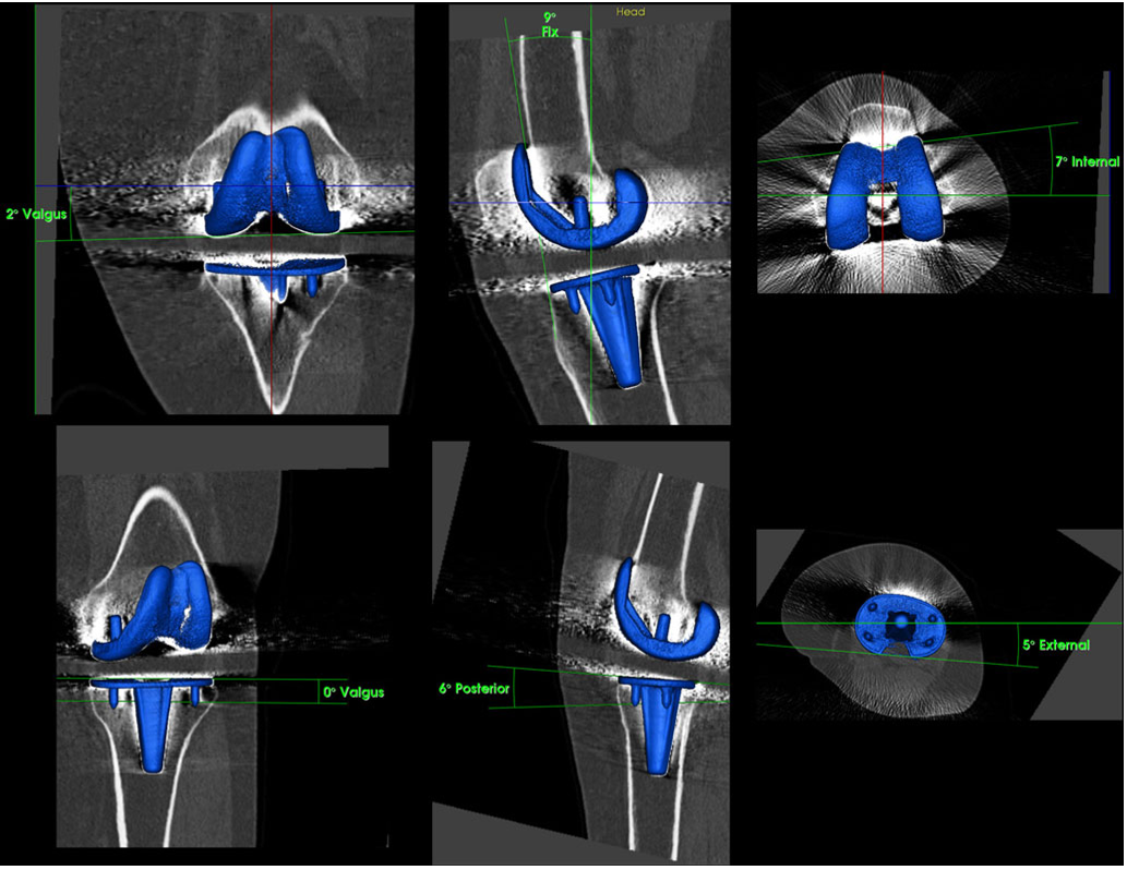
Fig. 2 Determination of tibial and femoral total knee arthroplasty
(TKA) component position (varus–valgus, flexion–extension, internal
rotation–external rotation) on 3D-CT using customised software after
reorientation in relation to the mechanical axis and definition of ana-
tomical landmarks on the bone surface (OrthoImagingSolutions,
London, UK)
Skeletal Radiol (2013) 42:1215–1223 1217

Measurements of TKA component position in 3D-CT
The position of the femoral and tibial TKA component was
assessed on 3D-CT after reorientation to the mechanical axis.
The sagittal (flexion–extension), coronal (varus–valgus) and
rotational alignment (internal rotation– external rotation) of
the femoral and tibial TKA components were determined on
3D-reconstructed CT images using customised software
(OrthoImagingSolutions, London, UK). The rotation of the
femoral component (femoral posterior component axis) was
measured in relation to the anatomical transepicondylar axis.
The rotation of the tibial component (tibial posterior compo-
nent axis) was measured in relation to the posterior tibial
plateau axis. One consultant radiologist/nuclear medicine spe-
cialist and one radiologist interpreted the SPECT/CTs for
tracer uptake and component analysis twice with a 2-week
interval between interpretations in a random order. Both were
blinded to results from previous observations.
All data were analysed by an independent professional
statistician using SPSS version 17.0 (SPSS, Chicago, IL,
USA.). Sample size was estimated according to the reported
estimates for reliability studies using intraclass correl ation
coefficients (ICCs) [17].
The inter- and intra-observer reliability of the intensity
and distribution analysis of SPECT/CT were determined by
calculating the intraclass correlation coefficients (ICC). An
ICC value of 1 indicated perfect reliability, 0.81 to 1 very
good reliability and 0.61 to 0.80 good reliability [17].
In addition, an inter-observer reliability analysis using the
Kappa statistic was performed to determine consistency
on the area with the highest tracer uptake. Kappa values
of < 0 represent poor, 0.0–0.20 slight, 0.21–0.40 fair, 0.41–
0.60 moderate, 0.61–0.80 substantial and 0.81–1.00 an
almost perfect agreement [18].
Results
All hotspots (areas with increased tracer uptake) were pres-
ent in the localisation scheme and could be located to
Fig. 3 The previously validated SPECT/CT scheme used for
localisation of the Tc-99m HDP tracer activity in patients with painful
knees after primary total knee arthroplasty. F femur, T tibia, P patella, 1
medial, 2 lateral, 3 central around stem, a anterior, p posterior, i
inferior, s superior, shaft, tip and tubercle. Reprinted with permission.
Publication can be found at www.springerlink.com [11]
1218 Skeletal Radiol (2013) 42:1215–1223

Table 1 Absolute tracer intensity values measured for each anatomical area and both observers
Observer 1 Observer 2 Both
Mean Standard deviation Median Minimum Maximum Mean Standard deviation Median Minimum Maximum Mean Standard deviation Median Minimum Maximum
f shaft 196.9 113.8 180 40 722 178.5 115.4 150 27 722 187.9 111.5 166 34 722
f med sa 472.4 389.1 381 47 2,550 442.8 383.8 364 38 2,550 457.7 384.3 379 43 2,550
f med sp 561.2 505.4 438 57 3,730 557.6 509.4 427 57 3,730 559.5 506.6 427 57 3,730
f med ia 441.9 308.7 360 31 1,652 427.3 303.1 353 30 1,652 434.8 303.2 367 31 1,652
f med ip 514.9 379.2 435 36 1,892 513.4 381.4 429 41 1,892 514.2 379.7 427 40 1,892
f lat sa 580.1 522.2 432 65 3,462 527.2 473.2 412 65 3,389 553.8 491.2 423 65 3,426
f lat sp 605.8 530.6 531 74 3,465 602.4 527.6 531 78 3,465 604.2 527.6 531 76 3,465
f lat ia 492.3 419.1 382 70 2,397 480.1 418.3 377 70 2,397 486.3 416.8 375 70 2,397
f lat ip 569.1 406.3 433 95 2,365 563.2 407.7 448 95 2,365 566.2 406 430 95 2,365
t med a 511.3 285.1 470 54 1,581 496.9 275.7 479 54 1,581 504.2 278.2 463 54 1,581
t med p 569.2 315.8 525 71 1,439 568.7 319.7 520 74 1,368 569 316.1 520 74 1,404
t lat a 483.3 275.3 460 77 1,496 488.5 277.2 457 77 1,496 485.9 274.3 460 77 1,496
t lat p 519.2 299.3 452 54 1,350 527.9 298.8 475 54 1,350 523.6 292.7 467 54 1,350
t stem 459.4 269.4 408 48 1,383 490.1 280.9 434 49 1,339 474.9 271.9 422 49 1,361
t tip 279.7 186.8 237 37 1,202 266.8 173.2 226 55 1,113 273.4 177.8 232 48 1,158
t shaft 214.2 113.5 189 46 552 161.1 82.8 146 34 378 187.9 93.8 174 41 428
p med s 591.8 386.5 512 77 1,852 597.4 398.4 502 77 1,852 594.7 390.7 508 77 1,852
p med i 527.7 333.2 500 61 1,715 521.6 347.9 483 61 1,775 524.8 336.9 490 61 1,715
p lat s 622.2 419.4 519 97 1,845 603.1 405.3 493 101 1,841 612.8 409.3 503 100 1,843
p lat i 566.6 394.1 463 103 1,602 552.8 396.3 440 98 1,760 559.9 390.7 442 103 1,602
p total 713.2 446.3 620 108 1,852 703.1 447.1 620 108 1,852 708.2 445.4 639 108 1,852
f femur, t tibia, p patella, sa superior–anterior, sp superior–posterior, ia inferior–anterior, ip inferior–posterior, med medial, lat lateral
Skeletal Radiol (2013) 42:1215–1223 1219

![Table 3 Intra- und inter-observer reliability (ICCs for absolute single-photon emission computed tomography [SPECT] values, relative SPECT values) of 3D voxel-based SPECT uptake measurement](/figures/table-3-intra-und-inter-observer-reliability-iccs-for-38xjzusw.png)


![Fig. 3 The previously validated SPECT/CT scheme used for localisation of the Tc-99m HDP tracer activity in patients with painful knees after primary total knee arthroplasty. F femur, T tibia, P patella, 1 medial, 2 lateral, 3 central around stem, a anterior, p posterior, i inferior, s superior, shaft, tip and tubercle. Reprinted with permission. Publication can be found at www.springerlink.com [11]](/figures/fig-3-the-previously-validated-spect-ct-scheme-used-for-3jq18chk.png)