scispace - formally typeset
Search or ask a question
Journal ArticleDOI

A bacteriophage detection tool for viability assessment of Salmonella cells.

TL;DR: This work presents and validates a novel bacteriophage (phage)-based microbial detection tool to detect and assess Salmonella viability and shows the phage selectivity in cell recognition minimizes false-negative and false-positive results often associated with most detection methods.
About: This article is published in Biosensors and Bioelectronics.The article was published on 2014-02-15 and is currently open access. It has received 90 citations till now. The article focuses on the topics: Bacteriophage.

Summary (3 min read)

1. Introduction

  • The ingestion of food, its derivatives and water contaminated with microbial pathogens (e.g. Escherichia coli, Campylobacter sp. or Salmonella sp.) is responsible for about 2.2 million deaths annually.
  • “Dormant” bacteria have therefore been called viable but non-culturable (VBNC) cells.
  • Significant progress has been reported in the phage-based detection of foodborne and waterborne pathogens (Hagens and Loessner, 2007; Singh et al., 2012; Smartt et al., 2012).

2.2. Bacteriophages and bacterial strains

  • PVP-SE1 was isolated from a Regensburg wastewater plant in the context of a European Project (Phagevet-P).
  • Salmonella Enteritidis strain S1400 was used as host (Sillankorva et al., 2010).
  • Campylobacter coli phage vB_CcoM-IBB_35, isolated from poultry intestines, was used as negative control (Carvalho et al., 2010a).

2.3. Phage propagation and buffer exchange

  • The phages were produced using the double layer agar technique as described by Sambrook and Russell (2001) and resuspended in SM buffer.
  • Exchange of SM buffer by MOPS buffer was needed to avoid the presence of amine groups from SM buffer, which may interfere with the surface chemistry adopted for phage immobilization on solid substrates.
  • Buffer exchange was made using a Vivaspin 500 centrifugal concentrator (MW 100 kDa).
  • Following the buffer exchange the concentration of phage was verified using the double layer agar technique.

2.4. Induction of Salmonella into viable but non-culturable (VBNC) state

  • Bacteria were induced to enter the VBNC state by using sodium hypochlorite (commercial bleach—stock concentration 5%) at different concentrations.
  • The serial dilutions of bleach were done with milli-Q water.
  • The samples were mixed at 200 rpm for 1 min at room temperature.
  • Following chlorination, the suspensions were centrifuged at 3420xg for 10 min at 4 1C and washed twice with cold PB.

2.5. Determination of cell viability

  • Cell viability was assessed after submitting bacteria to different bleach concentrations using the LIVE/DEADs BacLight™ Bacterial Viability and Counting Kit (Molecular Probes).
  • SYTO9 and PI dyes were used, accordingly to manufacturer's instructions.
  • Upon staining, cells were analyzed either by epifluorescence microscopy (OLYMPUS BX51 EXTREMO microscope) or by flow cytometry (BD LSRII flow cytometer using FACS DIVA software for acquisition; BD Biosciences).
  • For absolute cell quantification, 6 μm diameter microspheres were used at a known concentration in the flow cytometry acquisition.
  • Flow cytometry data was analyzed using the FlowJo software (Tree Star, Ashland, OR).

2.6. Phage lysis time and adsorption studies

  • 1 mL of each Salmonella sample was infected with PVP-SE1 phage at a multiplicity of infection (MOI) of 0.001, which refers to the number of phages that were added per cell.
  • Samples were taken immediately after infection (time 0) and after 20 min and 40 min of phage inoculation, followed by 10-fold dilution in MOPS and centrifugation at 10,000g for 10 min.
  • The supernatant was 10-fold serially-diluted in MOPS and plated to assess the concentration of PFU (plaque forming unit).
  • The phage adsorption fraction was calculated by dividing the PFU concentration at each time point by the initial phage concentration.
  • To assess the phage lysis time viable exponential phase grown Salmonella cells were used.

2.7. Phage immobilization on Au surfaces

  • Cr 5 nm/Au 40 nm thin film layers were sputtered (Kenosistec sputtering tool) over a silicon wafer.
  • The wafer was then spincoated with a photoresist (PR) polymer (AZ1505 AZ Electronic Materials) for surface protection and diced in 7 7 mm2 dies using an automatic dicing saw (Disco, DAD3350).
  • Substrates were then rinsed with isopropanol (IPA) and milli-Q water and dried under a nitrogen stream.
  • The gold surface was then functionalized with a heterobifunctional linker, the sulfo-LC-SPDP (sulfosuccinimidyl 6-[3′-(2-pyridyldithio)-propionamido] hexanoate).
  • Spot pictures were taken with an optical stereomicroscope (Nikon SMZ 1500) equipped with a CCD camera and analyzed using the image processing software ImageJ.

2.8. MR-biochip measurement

  • The MR-biochip was produced at INESC MN through a dedicated microfabrication process (Martins et al., 2009) and wirebonded to a PCB chip-carrier.
  • The probe sites on the MR biochip terminate with exposed Cr/Au pads, underneath which lie the magnetoresistive sensors that will detect the magnetic nanoparticle labels.
  • Briefly, the MR chip architecture comprises two distinct sensing areas arranged in two columns.
  • A 1 mL droplet of Salmonella-specific phage was spotted over the left column of sensors (12 sensors) and a non-specific phage (Campylobacter phage) on the right column of sensors (12 sensors).
  • The difference between the signal acquired after washing and the baseline signal is proportional to the number of cells bound to the sensor surface.

2.9. Antibody-conjugated MNPs preparation

  • Commercial 250 nm Protein A modified MNPs (Nanomag, Micromod) were used.
  • The unbound antibody was removed by the same magnetic separation procedure.
  • The functionalized MNPs were finally resuspended in 5 mL of PB Tw20 and injected over the chip.

2.10. Statistical analysis

  • All data are represented as mean7SD (standard deviation).
  • For Figs. 2 and 3, means were compared using two-way ANOVA followed by the Bonferroni post hoc test.

3.1. Induction of VBNC physiological state in Salmonella

  • Since the goal of this work was to prove the phage ability to detect the VBNC state of bacterial cells, a process was first developed capable of affecting cell viability in a controlled manner that would not lead to killing or lysing the entire cell population.
  • For this purpose different bactericidal and bacteriostatic compounds, known to induce the VBNC state in Salmonella cells, were tested (data not shown).
  • When exposed to fresh liquid medium under adequate growth conditions all tested concentrations of bleach, even above the break-point, showed cell growth (Supplementary data, Fig. S2.1).
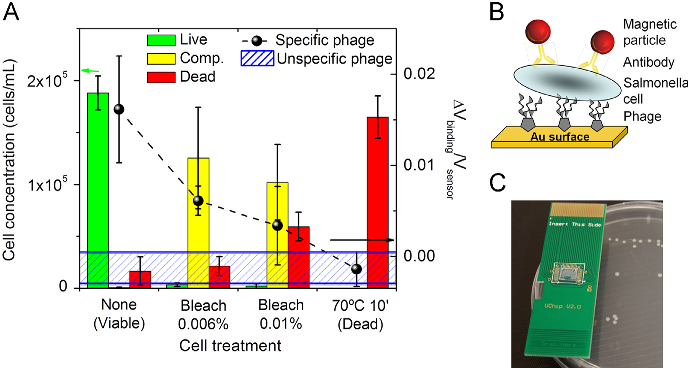
  • In order to quantitatively determine the relative and absolute proportion of the different cell populations (classified as live, dead or compromised), flow cytometry analyses were conducted for the different cell samples (Fig. 1A bars and 1C).
  • Results confirmed that, despite being present in sub-optimal host infection conditions, the phage adsorption capability was conserved, maintaining its potential to be used as a detection tool.

3.3. Phage performance as a biorecognition element

  • After optimization of the surface chemistry (Supplementary data, Fig. S3.1 and S3.2), the phage was immobilized on an Au surface at discrete areas by manual spotting.
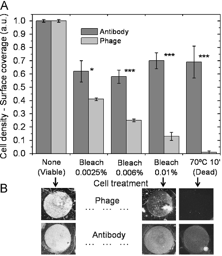
  • Also according to phage adsorption rates in solution, the immobilized phages were able to discriminate between viable and dead cells.
  • This resulted in reduced cell densities for samples with increasing number of dead cells (Fig. 3A) but proportional to the relative concentration of viable plus VBNC cells (compromised population) obtained by flow cytometry analysis (Fig. 1A—bars plot).
  • Identical biorecognition elements may hinder each other's proper attachment.
  • This is a common scenario in standard immunoassays where a labeling antibody may block the epitopes to the capture antibody or vice versa.

3.4. Phage-based magnetoresistive biochip for cell viability assessment

  • The feasibility of developing a “sandwich” phage-based biosensing system and its potential as a cell viability determination tool was assessed making use of an existent magnetoresistive (MR) biochip (Freitas et al., 2012; Martins et al., 2009, 2010) and respective electronic reader (Germano et al., 2009).
  • The biomolecular recognition strategy used on the biochip combines the phage and a magnetically-labeled antibody as recognition and labeling elements, respectively.
  • After the functionalization of the biochip with PVP-SE1 bacteriophage, each cell solution was loaded over the chip surface and incubated.
  • After washing, the magnetic fringe field created by the labels was detected as a variation on the sensor resistance.
  • Fig. 4A (dashed line and black dots) shows the biosensor normalized output for decreasing concentrations of viableþ VBNC cells.

4. Conclusions

  • The lytic phage PVP-SE1 was explored as an alternative biorecognition element for bacterial detection and viability assessment.
  • Taking into account the problematic occurrence of false positives associated with DNA-chips and the high production costs, poor stability and cross-reactivity related to immuno-chips, the development of phage-based biochips emerges as a valuable tool.
  • The feasibility to immobilize phages on sensing surfaces and conjugate this biomolecular tool with electronic analytical devices without losing functionality was proven.
  • The combined use of the magnetoresistive sensor with the phage probes allowed a clear detection of viable from dead Salmonella cells.

Did you find this useful? Give us your feedback

Figures (4)
Citations
More filters
Journal ArticleDOI
TL;DR: The results indicate that crRNAs selection is a major rate limiting factor in T4 phage engineering via CRISPR/Cas9 and the improved phage-engineering platform will accelerate the fields of individualized phage therapy, biocontrol, and rapid diagnostics.
Abstract: A major limitation hindering the widespread use of synthetic phages in medical and industrial settings is the lack of an efficient phage-engineering platform. Classical T4 phage engineering and several newly proposed methods are often inefficient and time consuming and consequently, only able to produce an inconsistent range of genomic editing rates between 0.03–3%. Here, we review and present new understandings of the CRISPR/Cas9 assisted genome engineering technique that significantly improves the genomic editing rate of T4 phages. Our results indicate that crRNAs selection is a major rate limiting factor in T4 phage engineering via CRISPR/Cas9. We were able to achieve an editing rate of > 99% for multiple genes that functionalizes the phages for further applications. We envision that this improved phage-engineering platform will accelerate the fields of individualized phage therapy, biocontrol, and rapid diagnostics.

29 citations

Journal ArticleDOI
TL;DR: This review comprehensively summarizes many uses of phages as detection tools and points the way toward how phage-based technologies may be improved.
Abstract: The innate specificity of bacteriophages toward their hosts makes them excellent candidates for the development of detection assays. They can be used in many ways to detect pathogens, and each has its own advantages and disadvantages. Whole bacteriophages can carry reporter genes to alter the phenotype of the target. Bacteriophages can act as staining agents or the progeny of the infection process can be detected, which further increases the sensitivity of the detection assay. Compared with whole-phage particles, use of phage components as probes offers other advantages: for example, smaller probe size to enhance binding activity, phage structures that can be engineered for better affinity, as well as specificity, binding properties, and robustness. When no natural binding with the target exists, phages can be used as vehicles to identify new protein-ligand interactions necessary for diagnostics. This review comprehensively summarizes many uses of phages as detection tools and points the way toward how phage-based technologies may be improved.

28 citations

Journal ArticleDOI
TL;DR: This work systematically investigated time- domain MRX by measuring the signal dependence on the applied field, magnetization time, and magnetic core size, and demonstrated, for the first time, a GMR-based time-domain MRX bioassay.
Abstract: Magnetorelaxometry (MRX) is a promising new biosensing technique for point-of-care diagnostics. Historically, magnetic sensors have been primarily used to monitor the stray field of magnetic nanoparticles bound to analytes of interest for immunoassays and flow cytometers. In MRX, the magnetic nanoparticles (MNPs) are first magnetized and then the temporal response is monitored after removing the magnetic field. This new sensing modality is insensitive to the magnetic field homogeneity making it more amenable to low-power portable applications. In this work, we systematically investigated time-domain MRX by measuring the signal dependence on the applied field, magnetization time, and magnetic core size. The extracted characteristic times varied for different magnetic MNPs, exhibiting unique magnetic signatures. We also measured the signal contribution based on the MNP location and correlated the coverage with measured signal amplitude. Lastly, we demonstrated, for the first time, a GMR-based time-domain MRX bioassay. This approach validates the feasibility of immunoassays using GMR-based MRX and provides an alternative platform for point-of-care diagnostics.

26 citations

Journal ArticleDOI
TL;DR: In this paper, a microfluidic system made of glass and polydimethylsiloxane was designed and fabricated for ex-vivo analysis of blood samples where superparamagnetic iron oxide nanoparticles, initially used as magnetic contrast agents, could be functionalized for biomarkers fishing.
Abstract: We have designed and fabricated a microfluidic system made of glass and polydimethylsiloxane. A micro-magnetometer has been integrated to the system. This sensor is made of a giant magneto-impedance wire known to have very high magnetic sensitivity at room temperature. A liquid-liquid segmented multiphase flow was generated in the channel using a Y-shaped inlet junction. The dispersed phase plugs contained superparamagnetic iron oxide (20 nm) nanoparticles at a molar concentration of 230 mmol/l. We have shown both theoretically and experimentally that in-flow detection of these nanoparticles is performed by the microsystem for concentration as small as 5.47 × 10⁻⁹ mol. These performances show that it is conceivable to use this system for ex-vivo analysis of blood samples where superparamagnetic iron oxide nanoparticles, initially used as magnetic contrast agents, could be functionalized for biomarkers fishing. It opens new perspectives in the context of personalized medicine.

25 citations

Journal ArticleDOI
TL;DR: In this paper, a single-layer graphene was modified with a heterobifunctional linker that attached to the surface via a pyrene group and presented a standard N-hydroxysuccinimide (NHS) ester ligand for covalent immobilization of biomolecules.

23 citations

References
More filters
Book
15 Jan 2001
TL;DR: Molecular Cloning has served as the foundation of technical expertise in labs worldwide for 30 years as mentioned in this paper and has been so popular, or so influential, that no other manual has been more widely used and influential.
Abstract: Molecular Cloning has served as the foundation of technical expertise in labs worldwide for 30 years. No other manual has been so popular, or so influential. Molecular Cloning, Fourth Edition, by the celebrated founding author Joe Sambrook and new co-author, the distinguished HHMI investigator Michael Green, preserves the highly praised detail and clarity of previous editions and includes specific chapters and protocols commissioned for the book from expert practitioners at Yale, U Mass, Rockefeller University, Texas Tech, Cold Spring Harbor Laboratory, Washington University, and other leading institutions. The theoretical and historical underpinnings of techniques are prominent features of the presentation throughout, information that does much to help trouble-shoot experimental problems. For the fourth edition of this classic work, the content has been entirely recast to include nucleic-acid based methods selected as the most widely used and valuable in molecular and cellular biology laboratories. Core chapters from the third edition have been revised to feature current strategies and approaches to the preparation and cloning of nucleic acids, gene transfer, and expression analysis. They are augmented by 12 new chapters which show how DNA, RNA, and proteins should be prepared, evaluated, and manipulated, and how data generation and analysis can be handled. The new content includes methods for studying interactions between cellular components, such as microarrays, next-generation sequencing technologies, RNA interference, and epigenetic analysis using DNA methylation techniques and chromatin immunoprecipitation. To make sense of the wealth of data produced by these techniques, a bioinformatics chapter describes the use of analytical tools for comparing sequences of genes and proteins and identifying common expression patterns among sets of genes. Building on thirty years of trust, reliability, and authority, the fourth edition of Mol
215,169 citations

Book
01 Jan 2001
TL;DR: The content has been entirely recast to include nucleic-acid based methods selected as the most widely used and valuable in molecular and cellular biology laboratories.
Abstract: Molecular Cloning has served as the foundation of technical expertise in labs worldwide for 30 years. No other manual has been so popular, or so influential. Molecular Cloning, Fourth Edition, by the celebrated founding author Joe Sambrook and new co-author, the distinguished HHMI investigator Michael Green, preserves the highly praised detail and clarity of previous editions and includes specific chapters and protocols commissioned for the book from expert practitioners at Yale, U Mass, Rockefeller University, Texas Tech, Cold Spring Harbor Laboratory, Washington University, and other leading institutions. The theoretical and historical underpinnings of techniques are prominent features of the presentation throughout, information that does much to help trouble-shoot experimental problems. For the fourth edition of this classic work, the content has been entirely recast to include nucleic-acid based methods selected as the most widely used and valuable in molecular and cellular biology laboratories. Core chapters from the third edition have been revised to feature current strategies and approaches to the preparation and cloning of nucleic acids, gene transfer, and expression analysis. They are augmented by 12 new chapters which show how DNA, RNA, and proteins should be prepared, evaluated, and manipulated, and how data generation and analysis can be handled. The new content includes methods for studying interactions between cellular components, such as microarrays, next-generation sequencing technologies, RNA interference, and epigenetic analysis using DNA methylation techniques and chromatin immunoprecipitation. To make sense of the wealth of data produced by these techniques, a bioinformatics chapter describes the use of analytical tools for comparing sequences of genes and proteins and identifying common expression patterns among sets of genes. Building on thirty years of trust, reliability, and authority, the fourth edition of Mol

25,596 citations


"A bacteriophage detection tool for ..." refers methods in this paper

  • ...For the lysis time studies, the bacterial cells were lysed and the total number of phages was quantified by the double layer agar technique (Sambrook and Russell, 2001)....

    [...]

  • ...The phages were produced using the double layer agar technique as described by Sambrook and Russell (2001) and resuspended in SM buffer....

    [...]

Journal Article
TL;DR: The ability of cells to resuscitate from the VBNC state and return to an actively metabolizing and culturable form is described, as well as the ability of these cells to retain virulence.
Abstract: It had long been assumed that a bacterial cell was dead when it was no longer able to grow on routine culture media. We now know that this assumption is simplistic, and that there are many situations where a cell loses culturability but remains viable and potentially able to regrow. This mini-review defines what the "viable but nonculturable" (VBNC) state is, and illustrates the methods that can be used to show that a bacterial cell is in this physiological state. The diverse environmental factors which induce this state, and the variety of bacteria which have been shown to enter into the VBNC state, are listed. In recent years, a great amount of research has revealed what occurs in cells as they enter and exist in this state, and these studies are also detailed. The ability of cells to resuscitate from the VBNC state and return to an actively metabolizing and culturable form is described, as well as the ability of these cells to retain virulence. Finally, the question of why cells become nonculturable is addressed. It is hoped that this mini-review will encourage researchers to consider this survival state in their studies as an alternative to the conclusion that a lack of culturability indicates the cells they are examining are dead.

1,237 citations


"A bacteriophage detection tool for ..." refers background in this paper

  • ...Nevertheless, they keep their virulence and the ability to resuscitate when in favorable conditions (Oliver, 2005, 2010)....

    [...]

Journal ArticleDOI
TL;DR: The central role of catalase in the VBNC response of some bacteria, including its genetic regulation, is described and a variety of interesting chemical and biological factors have been shown to allow resuscitation, including extracellular resuscitation-promoting proteins, a novel quorum-sensing system and interactions with amoeba.
Abstract: Many bacteria, including a variety of important human pathogens, are known to respond to various environmental stresses by entry into a novel physiological state, where the cells remain viable, but are no longer culturable on standard laboratory media. On resuscitation from this ‘viable but nonculturable’ (VBNC) state, the cells regain culturability and the renewed ability to cause infection. It is likely that the VBNC state is a survival strategy, although several interesting alternative explanations have been suggested. This review describes the VBNC state, the various chemical and physical factors known to induce cells into this state, the cellular traits and gene expression exhibited by VBNC cells, their antibiotic resistance, retention of virulence and ability to attach and persist in the environment, and factors that have been found to allow resuscitation of VBNC cells. Along with simple reversal of the inducing stresses, a variety of interesting chemical and biological factors have been shown to allow resuscitation, including extracellular resuscitation-promoting proteins, a novel quorum-sensing system (AI-3) and interactions with amoeba. Finally, the central role of catalase in the VBNC response of some bacteria, including its genetic regulation, is described.

1,029 citations


"A bacteriophage detection tool for ..." refers background in this paper

  • ...Nevertheless, they keep their virulence and the ability to resuscitate when in favorable conditions (Oliver, 2005, 2010)....

    [...]

Journal ArticleDOI
TL;DR: The conventional methods, analytical techniques and recent developments in food pathogen detection, identification and quantification, with an emphasis on biosensors are described.

1,023 citations


"A bacteriophage detection tool for ..." refers background in this paper

  • ...Such actions have stimulated R&D activities seeking for new methods for microbial detection, in particular bioanalytical technologies (Nugen and Baeumner, 2008; Velusamy et al., 2010)....

    [...]

Frequently Asked Questions (1)
Q1. What have the authors contributed in "A bacteriophage detection tool for viability assessment of salmonella cells" ?

This work presents and validates a novel bacteriophage ( phage ) -based microbial detection tool to detect and assess Salmonella viability. This ability was confirmed for immobilized phages on gold surfaces, where the phage detection signal follows the same trend of the concentration of viable plus VBNC cells in the sample. Salmonella Enteritidis cells in a VBNC physiological state were evaluated by cell culture, flow-cytometry and epifluorescence microscopy, and further assayed with a biosensor platform.