scispace - formally typeset
Search or ask a question
Journal ArticleDOI

A bacteriophage detection tool for viability assessment of Salmonella cells.

TL;DR: This work presents and validates a novel bacteriophage (phage)-based microbial detection tool to detect and assess Salmonella viability and shows the phage selectivity in cell recognition minimizes false-negative and false-positive results often associated with most detection methods.
About: This article is published in Biosensors and Bioelectronics.The article was published on 2014-02-15 and is currently open access. It has received 90 citations till now. The article focuses on the topics: Bacteriophage.

Summary (3 min read)

1. Introduction

  • The ingestion of food, its derivatives and water contaminated with microbial pathogens (e.g. Escherichia coli, Campylobacter sp. or Salmonella sp.) is responsible for about 2.2 million deaths annually.
  • “Dormant” bacteria have therefore been called viable but non-culturable (VBNC) cells.
  • Significant progress has been reported in the phage-based detection of foodborne and waterborne pathogens (Hagens and Loessner, 2007; Singh et al., 2012; Smartt et al., 2012).

2.2. Bacteriophages and bacterial strains

  • PVP-SE1 was isolated from a Regensburg wastewater plant in the context of a European Project (Phagevet-P).
  • Salmonella Enteritidis strain S1400 was used as host (Sillankorva et al., 2010).
  • Campylobacter coli phage vB_CcoM-IBB_35, isolated from poultry intestines, was used as negative control (Carvalho et al., 2010a).

2.3. Phage propagation and buffer exchange

  • The phages were produced using the double layer agar technique as described by Sambrook and Russell (2001) and resuspended in SM buffer.
  • Exchange of SM buffer by MOPS buffer was needed to avoid the presence of amine groups from SM buffer, which may interfere with the surface chemistry adopted for phage immobilization on solid substrates.
  • Buffer exchange was made using a Vivaspin 500 centrifugal concentrator (MW 100 kDa).
  • Following the buffer exchange the concentration of phage was verified using the double layer agar technique.

2.4. Induction of Salmonella into viable but non-culturable (VBNC) state

  • Bacteria were induced to enter the VBNC state by using sodium hypochlorite (commercial bleach—stock concentration 5%) at different concentrations.
  • The serial dilutions of bleach were done with milli-Q water.
  • The samples were mixed at 200 rpm for 1 min at room temperature.
  • Following chlorination, the suspensions were centrifuged at 3420xg for 10 min at 4 1C and washed twice with cold PB.

2.5. Determination of cell viability

  • Cell viability was assessed after submitting bacteria to different bleach concentrations using the LIVE/DEADs BacLight™ Bacterial Viability and Counting Kit (Molecular Probes).
  • SYTO9 and PI dyes were used, accordingly to manufacturer's instructions.
  • Upon staining, cells were analyzed either by epifluorescence microscopy (OLYMPUS BX51 EXTREMO microscope) or by flow cytometry (BD LSRII flow cytometer using FACS DIVA software for acquisition; BD Biosciences).
  • For absolute cell quantification, 6 μm diameter microspheres were used at a known concentration in the flow cytometry acquisition.
  • Flow cytometry data was analyzed using the FlowJo software (Tree Star, Ashland, OR).

2.6. Phage lysis time and adsorption studies

  • 1 mL of each Salmonella sample was infected with PVP-SE1 phage at a multiplicity of infection (MOI) of 0.001, which refers to the number of phages that were added per cell.
  • Samples were taken immediately after infection (time 0) and after 20 min and 40 min of phage inoculation, followed by 10-fold dilution in MOPS and centrifugation at 10,000g for 10 min.
  • The supernatant was 10-fold serially-diluted in MOPS and plated to assess the concentration of PFU (plaque forming unit).
  • The phage adsorption fraction was calculated by dividing the PFU concentration at each time point by the initial phage concentration.
  • To assess the phage lysis time viable exponential phase grown Salmonella cells were used.

2.7. Phage immobilization on Au surfaces

  • Cr 5 nm/Au 40 nm thin film layers were sputtered (Kenosistec sputtering tool) over a silicon wafer.
  • The wafer was then spincoated with a photoresist (PR) polymer (AZ1505 AZ Electronic Materials) for surface protection and diced in 7 7 mm2 dies using an automatic dicing saw (Disco, DAD3350).
  • Substrates were then rinsed with isopropanol (IPA) and milli-Q water and dried under a nitrogen stream.
  • The gold surface was then functionalized with a heterobifunctional linker, the sulfo-LC-SPDP (sulfosuccinimidyl 6-[3′-(2-pyridyldithio)-propionamido] hexanoate).
  • Spot pictures were taken with an optical stereomicroscope (Nikon SMZ 1500) equipped with a CCD camera and analyzed using the image processing software ImageJ.

2.8. MR-biochip measurement

  • The MR-biochip was produced at INESC MN through a dedicated microfabrication process (Martins et al., 2009) and wirebonded to a PCB chip-carrier.
  • The probe sites on the MR biochip terminate with exposed Cr/Au pads, underneath which lie the magnetoresistive sensors that will detect the magnetic nanoparticle labels.
  • Briefly, the MR chip architecture comprises two distinct sensing areas arranged in two columns.
  • A 1 mL droplet of Salmonella-specific phage was spotted over the left column of sensors (12 sensors) and a non-specific phage (Campylobacter phage) on the right column of sensors (12 sensors).
  • The difference between the signal acquired after washing and the baseline signal is proportional to the number of cells bound to the sensor surface.

2.9. Antibody-conjugated MNPs preparation

  • Commercial 250 nm Protein A modified MNPs (Nanomag, Micromod) were used.
  • The unbound antibody was removed by the same magnetic separation procedure.
  • The functionalized MNPs were finally resuspended in 5 mL of PB Tw20 and injected over the chip.

2.10. Statistical analysis

  • All data are represented as mean7SD (standard deviation).
  • For Figs. 2 and 3, means were compared using two-way ANOVA followed by the Bonferroni post hoc test.

3.1. Induction of VBNC physiological state in Salmonella

  • Since the goal of this work was to prove the phage ability to detect the VBNC state of bacterial cells, a process was first developed capable of affecting cell viability in a controlled manner that would not lead to killing or lysing the entire cell population.
  • For this purpose different bactericidal and bacteriostatic compounds, known to induce the VBNC state in Salmonella cells, were tested (data not shown).
  • When exposed to fresh liquid medium under adequate growth conditions all tested concentrations of bleach, even above the break-point, showed cell growth (Supplementary data, Fig. S2.1).
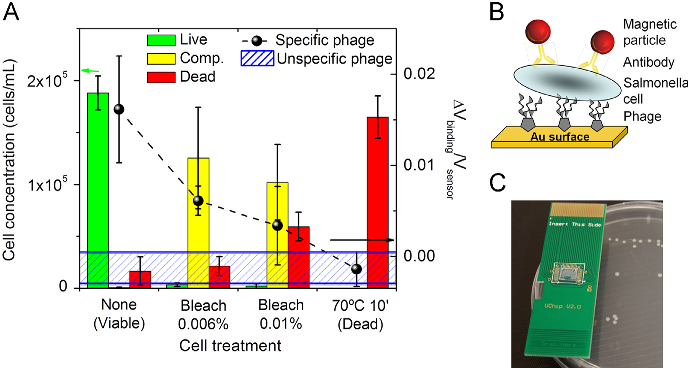
  • In order to quantitatively determine the relative and absolute proportion of the different cell populations (classified as live, dead or compromised), flow cytometry analyses were conducted for the different cell samples (Fig. 1A bars and 1C).
  • Results confirmed that, despite being present in sub-optimal host infection conditions, the phage adsorption capability was conserved, maintaining its potential to be used as a detection tool.

3.3. Phage performance as a biorecognition element

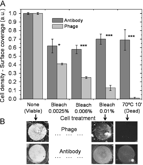
  • After optimization of the surface chemistry (Supplementary data, Fig. S3.1 and S3.2), the phage was immobilized on an Au surface at discrete areas by manual spotting.
  • Also according to phage adsorption rates in solution, the immobilized phages were able to discriminate between viable and dead cells.
  • This resulted in reduced cell densities for samples with increasing number of dead cells (Fig. 3A) but proportional to the relative concentration of viable plus VBNC cells (compromised population) obtained by flow cytometry analysis (Fig. 1A—bars plot).
  • Identical biorecognition elements may hinder each other's proper attachment.
  • This is a common scenario in standard immunoassays where a labeling antibody may block the epitopes to the capture antibody or vice versa.

3.4. Phage-based magnetoresistive biochip for cell viability assessment

  • The feasibility of developing a “sandwich” phage-based biosensing system and its potential as a cell viability determination tool was assessed making use of an existent magnetoresistive (MR) biochip (Freitas et al., 2012; Martins et al., 2009, 2010) and respective electronic reader (Germano et al., 2009).
  • The biomolecular recognition strategy used on the biochip combines the phage and a magnetically-labeled antibody as recognition and labeling elements, respectively.
  • After the functionalization of the biochip with PVP-SE1 bacteriophage, each cell solution was loaded over the chip surface and incubated.
  • After washing, the magnetic fringe field created by the labels was detected as a variation on the sensor resistance.
  • Fig. 4A (dashed line and black dots) shows the biosensor normalized output for decreasing concentrations of viableþ VBNC cells.

4. Conclusions

  • The lytic phage PVP-SE1 was explored as an alternative biorecognition element for bacterial detection and viability assessment.
  • Taking into account the problematic occurrence of false positives associated with DNA-chips and the high production costs, poor stability and cross-reactivity related to immuno-chips, the development of phage-based biochips emerges as a valuable tool.
  • The feasibility to immobilize phages on sensing surfaces and conjugate this biomolecular tool with electronic analytical devices without losing functionality was proven.
  • The combined use of the magnetoresistive sensor with the phage probes allowed a clear detection of viable from dead Salmonella cells.

Did you find this useful? Give us your feedback

Figures (4)
Citations
More filters
Journal ArticleDOI
TL;DR: Various aspects of VBNC bacteria are described, which include their proteomic and genetic profiles under the VB NC state, conditions of resuscitation, methods of detection, antibiotic resistance, and observations on Rpf.
Abstract: Under stress conditions, many species of bacteria enter into starvation mode of metabolism or a physiologically viable but non-culturable (VBNC) state. Several human pathogenic bacteria have been reported to enter into the VBNC state under these conditions. The pathogenic VBNC bacteria cannot be grown using conventional culture media, although they continue to retain their viability and express their virulence. Though there have been debates on the VBNC concept in the past, several molecular studies have shown that not only can the VBNC state be induced under in vitro conditions but also that resuscitation from this state is possible under appropriate conditions. The most notable advance in resuscitating VBNC bacteria is the discovery of resuscitation-promoting factor (Rpf), which is a bacterial cytokines found in both Gram-positive and Gram-negative organisms. VBNC state is a survival strategy adopted by the bacteria, which has important implication in several fields, including environmental monitoring, food technology, and infectious disease management; and hence it is important to investigate the association of bacterial pathogens under VBNC state and the water/foodborne outbreaks. In this review, we describe various aspects of VBNC bacteria, which include their proteomic and genetic profiles under the VBNC state, conditions of resuscitation, methods of detection, antibiotic resistance, and observations on Rpf.

338 citations


Cites background from "A bacteriophage detection tool for ..."

  • ...” Several recent studies have shown the usefulness of bacteriophages in the detection of VBNC cells present in the bacterial populations (72, 73)....

    [...]

Journal ArticleDOI
TL;DR: This review highlights advances in techniques used to engineer phages as vehicles for drug delivery and vaccines, as well as for the assembly of new materials, and discusses existing challenges and opportunities.
Abstract: Soon after their discovery in the early 20th century, bacteriophages were recognized to have great potential as antimicrobial agents, a potential that has yet to be fully realized. The nascent field of phage therapy was adversely affected by inadequately controlled trials and the discovery of antibiotics. Although the study of phages as anti-infective agents slowed, phages played an important role in the development of molecular biology. In recent years, the increase in multidrug-resistant bacteria has renewed interest in the use of phages as antimicrobial agents. With the wide array of possibilities offered by genetic engineering, these bacterial viruses are being modified to precisely control and detect bacteria and to serve as new sources of antibacterials. In applications that go beyond their antimicrobial activity, phages are also being developed as vehicles for drug delivery and vaccines, as well as for the assembly of new materials. This review highlights advances in techniques used to engineer phages for all of these purposes and discusses existing challenges and opportunities for future work.

295 citations

Journal ArticleDOI
TL;DR: This review provides an overview of the biology of the VB NC state, its relationship to food safety, and novel methods developed for the rapid detection and identification of VBNC cells.
Abstract: The viable but non-culturable (VBNC) state is a form of dormancy employed by many bacteria as a method of survival and can be found in nearly any ecological niche. Major characteristics that distinguish dormant cells is their ability to evade detection by routine laboratory culture, to tolerate stressful environments including food pasteurization processes and antibiotics, and to resuscitate within a host and cause disease. Given these defining characteristics, these resilient microbes raise significant concern for the food industry and for the health of those consuming foods harboring these veiled pathogens. This review provides an overview of the biology of the VBNC state, its relationship to food safety, and novel methods developed for the rapid detection and identification of VBNC cells.

97 citations

Journal ArticleDOI
TL;DR: The importance of electrochemical biosensors as simple, reliable, cost-effective, and accurate tools for bacterial detection is emphasized, as well as the most recent advancements in phage-based sensing assays and sensors.

89 citations

Journal ArticleDOI
TL;DR: This review proposes to gather and comment different ligands used for the detection of whole cell bacteria and label-free methods, which enable the user to skip sampling processing steps and decrease the overall test cost.
Abstract: With the aim of getting earlier, sensitive and specific information on the presence –or absence – of bacterial pathogens, biosensors are getting an increasing interest for more than two decades. This is partly due to their reduced format, to the possibility to address several questions with a single device and also to the increasing panel of physical approaches that can be exploited for signal transducing. When designing a biosensor, the choice of the ligand motif remains a key element as it drives the efficiency and sensitivity of the assay. In this review, we propose to gather and comment different ligands used for the detection of whole cell bacteria. Because time is a crucial issue when looking for a pathogen, our attention was focused on whole cell assays and label-free methods, which enable the user to skip sampling processing steps and decrease the overall test cost.

89 citations

References
More filters
Journal ArticleDOI
TL;DR: Late-exponential-phase cells of Escherichia coli O157:H- strain E32511/HSC became nonculturable in sterilized distilled water microcosms at 4 °C because of starvation and a low temperature and the proposed mode of action of the catalase or pyruvate is via the degradation of the metabolic by-product H2O2.
Abstract: Late-exponential-phase cells of Escherichia coli O157:H- strain E32511/HSC became nonculturable in sterilized distilled water microcosms at 4 degrees C. Plate counts declined from 3 x 10(6) to less than 0.1 CFU/ml in about 21 days. However, when samples of microcosms at 21 days were inoculated onto an agar medium amended with catalase or nonenzyme peroxide-degrading compounds such as sodium pyruvate or alpha-ketoglutaric acid, plate counts increased to 10(4)-10(5) CFU/ml within 48 h. The proposed mode of action of the catalase or pyruvate is via the degradation of the metabolic by-product H2O2, rather than through supplementation of a required nutrient in the recovery of nonculturable cells. Our studies were based on the assumption that E32511/HSC strain responds to starvation and a low temperature by entering a nonculturable state and that the correction of oxidative stress upon the inoculation of bacteria on agar plates promotes recovery of nonculturable cells.

178 citations


"A bacteriophage detection tool for ..." refers background in this paper

  • ...The VBNC state has been associated to a survival mechanism of the bacteria upon exposure to harsh but sub-lethal conditions (Mizunoe et al., 1999)....

    [...]

Journal ArticleDOI
TL;DR: In this article, a microfluidic chip utilizes impedance-based measurement to detect cells and identify them when used in conjunction with immobilized monoclonal antibodies, which increases the measured impedance within the chamber.
Abstract: In this paper, we have developed a simple and rapid method for the detection and identification of bacteria using a microfluidic lab-chip. The microfluidic chip utilizes impedance-based measurement to (1) detect cells and (2) identify them when used in conjunction with immobilized monoclonal antibodies. Bacteria in suspension passing through the microfluidic chamber are recognized by antibodies and selectively immobilized on the functionalized glass surface, thereby increasing the measured impedance within the chamber. Continuous perfusion of bacteria suspension through the derivatized chamber not only identifies specific bacteria but also enhances the chamber's detection sensitivity by accumulating bacteria on the chamber wall over time; this approach would be useful for detecting low concentrations of bacteria. To demonstrate this approach, we showed that the prototype sensor could detect 9 × 10⁵ CFU mL−1 E. coli (BL21(DE3)) in the solution by consecutive perfusions. The chip sensitivity with immobilized bacteria is governed by height of sensing chamber, and ∼104 CFU mL−1 of E. coli could easily be detected when a shallower chamber (2 μm high) was used. The selectivity of the sensor was tested using a suspension of two bacterial strains, E. coli and M. catarrhalis. The sensor chip is simple to use, requires minuscule samples, and eliminates extensive cell culture processes. Development of more advanced lab-chips with multiple chambers containing different antibodies that allow simultaneous detection of different bacteria strains will be a natural extension of this work.

174 citations


"A bacteriophage detection tool for ..." refers result in this paper

  • ...In contrast to the actual culture-based methods, biosensors have started to offer great advantages due to their faster and more sensitive response (Boehm et al., 2007; Ivnitski et al., 1999)....

    [...]

Journal ArticleDOI
TL;DR: In this paper, a triple fluorochrome staining procedure was developed that takes account of the problems of active dye extrusion or cell dormancy on viability measurements used to date (e.g., enzyme activity or cell polarization).
Abstract: Rapid bacterial detection and viability measurements have been greatly enhanced by recent advances in the use of fluorescent stains in cytometry. It has previously been shown that four physiological states can be distinguished: reproductively viable, metabolically active, intact and permeabilized. Previous sorting experiments have shown that not all intact cells readily grow, but some intact cells can grow even when they fail to show metabolic activity, as determined by esterase turnover. To circumvent the limitations imposed by active dye extrusion or cell dormancy on viability measurements used to date (e.g., enzyme activity or cell polarization), a fast triple fluorochrome staining procedure has been developed that takes account of these problems. This allows further cellular characterization of intact cells by: active exclusion of ethidium bromide (EB) (metabolically active cells), uptake of EB but exclusion of bis-oxonol (BOX) (de-energized but with a polarized cell membrane) and uptake of both dyes (depolarized). Permeabilized cells were identified by propidium iodide (PI) uptake. The method was validated using an electronically programmable single cell sorter (EPICS Elite) and aged Salmonella typhimurium cells. Reproductive viability was determined by sorting single cells to their staining pattern directly onto agar plates. Most polarized cells could be recovered as well as a significant fraction of the depolarized cells, demonstrating that depolarization is a sensitive measure of cell damage but a poor indicator of cell death.

170 citations

Journal ArticleDOI
TL;DR: Food safety practices have vastly improved in the processing environment as a result of these regulatory actions, as evidenced by the decreasing rates of infection by both Listeria and E. coli O157:H7.
Abstract: Despite the recent advances in food pathogen detection, there still exist many challenges and opportunities to improve the current technology. Techniques such as immunomagnetic separation (IMS) and polymerase chain reaction (PCR) have paved the way for rapid and sensitive detection of foodborne pathogens, and advances in nanobiotechnology have allowed for miniaturization of devices. Collaborations between workers in the fields of engineering, nanotechnology and food science have introduced new lab-on-a-chip technologies permitting development of portable, hand-held biosensors for food pathogen detection. This report highlights examples within the current state of the art, and emphasizes areas in which further research is needed. In 1999 it was estimated that foodborne pathogens were responsible for 76 million illnesses annually, resulting in 5,000 deaths [1]. This report identified Salmonella, Listeria and Toxoplasma as the major causative agents, being responsible for 1,500 of the reported deaths. Data published in 2006 by the CDC suggested that infections due to Yersinia, Shigella, Listeria, Campylobacter, Escherichia coli O157:H7 and Salmonella have decreased dramatically, while infections due to Vibrio have increased [2]. A more recent report indicated similar findings, with a decrease in Yersinia, Shigella, Listeria and Campylobacter cases, and again a significant increase in Vibrio infections [3]. The declining rates of infection due to Listeria monocytogenes and E. coli O157:H7 are likely a result of increased awareness. The FDA, USDA and EU have all implemented a zero-tolerance rule for L. monocytogenes in ready-to-eat (RTE) foods. Similarly, the USDA’s Food Safety and Inspection Service (USDA-FSIS) has declared E. coli O157:H7 in raw ground beef to be an adulterant and therefore unfit for human consumption. Food safety practices have vastly improved in the processing environment as a result of these regulatory actions, as evidenced by the decreasing rates of infection by both Listeria and E. coli O157:H7. However, of the 121 foodborne outbreaks reported through FoodNet in 2005, almost half (49%) of the reported cases were attributed to noroviruses, and the number of Vibrio infections is on the rise [2]. Therefore, while the “hot” organism may change and while food safety practices are improving, there remains a growing need for enhanced means of food pathogen detection. In addition to the health risk associated with contaminated foods, there is the often devastating economic impact to the food producer. A 2007 recall of 21. 7 × 10⁶ lb of ground beef owing to contamination with E. coli O157:H7 resulted in the Topps Meat Company going out of business after 67 years of operation. Indeed, the consolidation of food producers in the USA means that the larger companies have a greater responsibility toward protecting the food supply. Coast-to-coast and international distribution by these mega-processing plants puts potential outbreaks on a national and international scale. Increasing automation in food processing facilities increases the risk of contamination by environmental sources following heat treatment, which is a critical concern especially for RTE products. Therefore, monitoring of pathogen counts on processing surfaces is critical in maintaining low or zero counts in food products. The costs of warehousing along with the potential costs of product recalls have potentially made on-site pathogen testing economically advantageous. Faster results would mean that products could go to market earlier. Several companies such as Marshfield Food Safety and IEH now offer on-site testing services for food production facilities. The latter can equip a production facility with modular laboratories that can be brought in, providing faster turnaround results. These needs stem from the increasing number of microbiological tests commissioned for analysis each week. A 2000 study estimated the number of weekly tests commissioned per dairy plant to be 636, followed closely by 444 weekly microbiological tests from processed-food plants [4].
144 citations


"A bacteriophage detection tool for ..." refers background in this paper

  • ...Such actions have stimulated R&D activities seeking for new methods for microbial detection, in particular bioanalytical technologies (Nugen and Baeumner, 2008; Velusamy et al., 2010)....

    [...]

Journal ArticleDOI
TL;DR: The GFP-labeled PP01 phage could be a rapid and sensitive tool for E. coli O157:H7 detection and increase the binding affinity of the recombinant phages to the host cell increased, however, the stability of the reunification phages in alkaline solution decreased.
Abstract: A previously isolated T-even-type PP01 bacteriophage was used to detect its host cell, Escherichia coli O157:H7. The phage small outer capsid (SOC) protein was used as a platform to present a marker protein, green fluorescent protein (GFP), on the phage capsid. The DNA fragment around soc was amplified by PCR and sequenced. The gene alignment of soc and its upstream region was g56-soc.2-soc.1-soc, which is the same as that for T2 phage. GFP was introduced into the C- and N-terminal regions of SOC to produce recombinant phages PP01-GFP/SOC and PP01-SOC/GFP, respectively. Fusion of GFP to SOC did not change the host range of PP01. On the contrary, the binding affinity of the recombinant phages to the host cell increased. However, the stability of the recombinant phages in alkaline solution decreased. Adsorption of the GFP-labeled PP01 phages to the E. coli cell surface enabled visualization of cells under a fluorescence microscope. GFP-labeled PP01 phage was not only adsorbed on culturable E. coli cells but also on viable but nonculturable or pasteurized cells. The coexistence of insensitive E. coli K-12 (W3110) cells did not influence the specificity and affinity of GFP-labeled PP01 adsorption on E. coli O157:H7. After a 10-min incubation with GFP-labeled PP01 phage at a multiplicity of infection of 1,000 at 4°C, E. coli O157:H7 cells could be visualized by fluorescence microscopy. The GFP-labeled PP01 phage could be a rapid and sensitive tool for E. coli O157:H7 detection.

143 citations


"A bacteriophage detection tool for ..." refers background in this paper

  • ...Studies on the utilization of phages for the detection of VBNC bacteria are still limited and have been applied only to E. coli O157:H7 (Awais et al., 2006; Oda et al., 2004)....

    [...]

Frequently Asked Questions (1)
Q1. What have the authors contributed in "A bacteriophage detection tool for viability assessment of salmonella cells" ?

This work presents and validates a novel bacteriophage ( phage ) -based microbial detection tool to detect and assess Salmonella viability. This ability was confirmed for immobilized phages on gold surfaces, where the phage detection signal follows the same trend of the concentration of viable plus VBNC cells in the sample. Salmonella Enteritidis cells in a VBNC physiological state were evaluated by cell culture, flow-cytometry and epifluorescence microscopy, and further assayed with a biosensor platform.