scispace - formally typeset
Search or ask a question
Journal ArticleDOI

A bacteriophage detection tool for viability assessment of Salmonella cells.

TL;DR: This work presents and validates a novel bacteriophage (phage)-based microbial detection tool to detect and assess Salmonella viability and shows the phage selectivity in cell recognition minimizes false-negative and false-positive results often associated with most detection methods.
About: This article is published in Biosensors and Bioelectronics.The article was published on 2014-02-15 and is currently open access. It has received 90 citations till now. The article focuses on the topics: Bacteriophage.

Summary (3 min read)

1. Introduction

  • The ingestion of food, its derivatives and water contaminated with microbial pathogens (e.g. Escherichia coli, Campylobacter sp. or Salmonella sp.) is responsible for about 2.2 million deaths annually.
  • “Dormant” bacteria have therefore been called viable but non-culturable (VBNC) cells.
  • Significant progress has been reported in the phage-based detection of foodborne and waterborne pathogens (Hagens and Loessner, 2007; Singh et al., 2012; Smartt et al., 2012).

2.2. Bacteriophages and bacterial strains

  • PVP-SE1 was isolated from a Regensburg wastewater plant in the context of a European Project (Phagevet-P).
  • Salmonella Enteritidis strain S1400 was used as host (Sillankorva et al., 2010).
  • Campylobacter coli phage vB_CcoM-IBB_35, isolated from poultry intestines, was used as negative control (Carvalho et al., 2010a).

2.3. Phage propagation and buffer exchange

  • The phages were produced using the double layer agar technique as described by Sambrook and Russell (2001) and resuspended in SM buffer.
  • Exchange of SM buffer by MOPS buffer was needed to avoid the presence of amine groups from SM buffer, which may interfere with the surface chemistry adopted for phage immobilization on solid substrates.
  • Buffer exchange was made using a Vivaspin 500 centrifugal concentrator (MW 100 kDa).
  • Following the buffer exchange the concentration of phage was verified using the double layer agar technique.

2.4. Induction of Salmonella into viable but non-culturable (VBNC) state

  • Bacteria were induced to enter the VBNC state by using sodium hypochlorite (commercial bleach—stock concentration 5%) at different concentrations.
  • The serial dilutions of bleach were done with milli-Q water.
  • The samples were mixed at 200 rpm for 1 min at room temperature.
  • Following chlorination, the suspensions were centrifuged at 3420xg for 10 min at 4 1C and washed twice with cold PB.

2.5. Determination of cell viability

  • Cell viability was assessed after submitting bacteria to different bleach concentrations using the LIVE/DEADs BacLight™ Bacterial Viability and Counting Kit (Molecular Probes).
  • SYTO9 and PI dyes were used, accordingly to manufacturer's instructions.
  • Upon staining, cells were analyzed either by epifluorescence microscopy (OLYMPUS BX51 EXTREMO microscope) or by flow cytometry (BD LSRII flow cytometer using FACS DIVA software for acquisition; BD Biosciences).
  • For absolute cell quantification, 6 μm diameter microspheres were used at a known concentration in the flow cytometry acquisition.
  • Flow cytometry data was analyzed using the FlowJo software (Tree Star, Ashland, OR).

2.6. Phage lysis time and adsorption studies

  • 1 mL of each Salmonella sample was infected with PVP-SE1 phage at a multiplicity of infection (MOI) of 0.001, which refers to the number of phages that were added per cell.
  • Samples were taken immediately after infection (time 0) and after 20 min and 40 min of phage inoculation, followed by 10-fold dilution in MOPS and centrifugation at 10,000g for 10 min.
  • The supernatant was 10-fold serially-diluted in MOPS and plated to assess the concentration of PFU (plaque forming unit).
  • The phage adsorption fraction was calculated by dividing the PFU concentration at each time point by the initial phage concentration.
  • To assess the phage lysis time viable exponential phase grown Salmonella cells were used.

2.7. Phage immobilization on Au surfaces

  • Cr 5 nm/Au 40 nm thin film layers were sputtered (Kenosistec sputtering tool) over a silicon wafer.
  • The wafer was then spincoated with a photoresist (PR) polymer (AZ1505 AZ Electronic Materials) for surface protection and diced in 7 7 mm2 dies using an automatic dicing saw (Disco, DAD3350).
  • Substrates were then rinsed with isopropanol (IPA) and milli-Q water and dried under a nitrogen stream.
  • The gold surface was then functionalized with a heterobifunctional linker, the sulfo-LC-SPDP (sulfosuccinimidyl 6-[3′-(2-pyridyldithio)-propionamido] hexanoate).
  • Spot pictures were taken with an optical stereomicroscope (Nikon SMZ 1500) equipped with a CCD camera and analyzed using the image processing software ImageJ.

2.8. MR-biochip measurement

  • The MR-biochip was produced at INESC MN through a dedicated microfabrication process (Martins et al., 2009) and wirebonded to a PCB chip-carrier.
  • The probe sites on the MR biochip terminate with exposed Cr/Au pads, underneath which lie the magnetoresistive sensors that will detect the magnetic nanoparticle labels.
  • Briefly, the MR chip architecture comprises two distinct sensing areas arranged in two columns.
  • A 1 mL droplet of Salmonella-specific phage was spotted over the left column of sensors (12 sensors) and a non-specific phage (Campylobacter phage) on the right column of sensors (12 sensors).
  • The difference between the signal acquired after washing and the baseline signal is proportional to the number of cells bound to the sensor surface.

2.9. Antibody-conjugated MNPs preparation

  • Commercial 250 nm Protein A modified MNPs (Nanomag, Micromod) were used.
  • The unbound antibody was removed by the same magnetic separation procedure.
  • The functionalized MNPs were finally resuspended in 5 mL of PB Tw20 and injected over the chip.

2.10. Statistical analysis

  • All data are represented as mean7SD (standard deviation).
  • For Figs. 2 and 3, means were compared using two-way ANOVA followed by the Bonferroni post hoc test.

3.1. Induction of VBNC physiological state in Salmonella

  • Since the goal of this work was to prove the phage ability to detect the VBNC state of bacterial cells, a process was first developed capable of affecting cell viability in a controlled manner that would not lead to killing or lysing the entire cell population.
  • For this purpose different bactericidal and bacteriostatic compounds, known to induce the VBNC state in Salmonella cells, were tested (data not shown).
  • When exposed to fresh liquid medium under adequate growth conditions all tested concentrations of bleach, even above the break-point, showed cell growth (Supplementary data, Fig. S2.1).
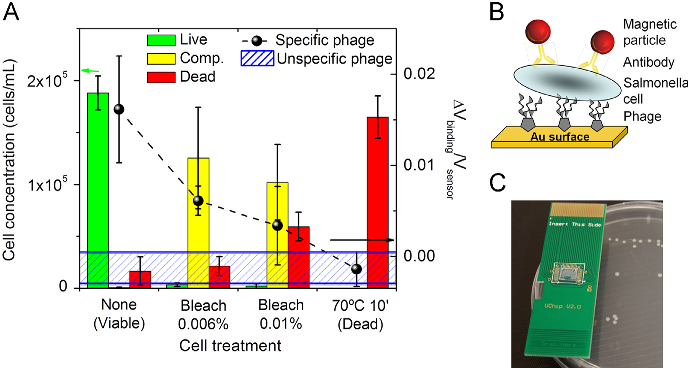
  • In order to quantitatively determine the relative and absolute proportion of the different cell populations (classified as live, dead or compromised), flow cytometry analyses were conducted for the different cell samples (Fig. 1A bars and 1C).
  • Results confirmed that, despite being present in sub-optimal host infection conditions, the phage adsorption capability was conserved, maintaining its potential to be used as a detection tool.

3.3. Phage performance as a biorecognition element

  • After optimization of the surface chemistry (Supplementary data, Fig. S3.1 and S3.2), the phage was immobilized on an Au surface at discrete areas by manual spotting.
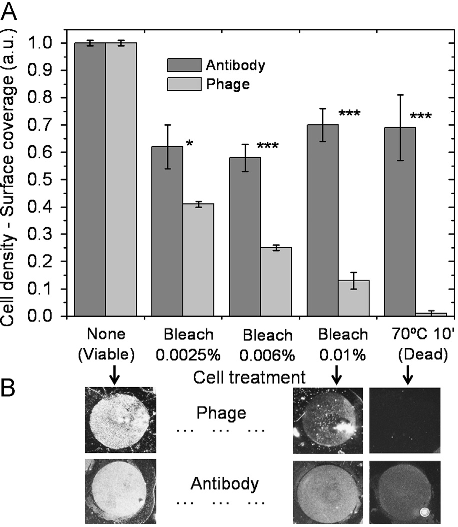
  • Also according to phage adsorption rates in solution, the immobilized phages were able to discriminate between viable and dead cells.
  • This resulted in reduced cell densities for samples with increasing number of dead cells (Fig. 3A) but proportional to the relative concentration of viable plus VBNC cells (compromised population) obtained by flow cytometry analysis (Fig. 1A—bars plot).
  • Identical biorecognition elements may hinder each other's proper attachment.
  • This is a common scenario in standard immunoassays where a labeling antibody may block the epitopes to the capture antibody or vice versa.

3.4. Phage-based magnetoresistive biochip for cell viability assessment

  • The feasibility of developing a “sandwich” phage-based biosensing system and its potential as a cell viability determination tool was assessed making use of an existent magnetoresistive (MR) biochip (Freitas et al., 2012; Martins et al., 2009, 2010) and respective electronic reader (Germano et al., 2009).
  • The biomolecular recognition strategy used on the biochip combines the phage and a magnetically-labeled antibody as recognition and labeling elements, respectively.
  • After the functionalization of the biochip with PVP-SE1 bacteriophage, each cell solution was loaded over the chip surface and incubated.
  • After washing, the magnetic fringe field created by the labels was detected as a variation on the sensor resistance.
  • Fig. 4A (dashed line and black dots) shows the biosensor normalized output for decreasing concentrations of viableþ VBNC cells.

4. Conclusions

  • The lytic phage PVP-SE1 was explored as an alternative biorecognition element for bacterial detection and viability assessment.
  • Taking into account the problematic occurrence of false positives associated with DNA-chips and the high production costs, poor stability and cross-reactivity related to immuno-chips, the development of phage-based biochips emerges as a valuable tool.
  • The feasibility to immobilize phages on sensing surfaces and conjugate this biomolecular tool with electronic analytical devices without losing functionality was proven.
  • The combined use of the magnetoresistive sensor with the phage probes allowed a clear detection of viable from dead Salmonella cells.

Did you find this useful? Give us your feedback

Figures (4)
Citations
More filters
Journal ArticleDOI
TL;DR: Various aspects of VBNC bacteria are described, which include their proteomic and genetic profiles under the VB NC state, conditions of resuscitation, methods of detection, antibiotic resistance, and observations on Rpf.
Abstract: Under stress conditions, many species of bacteria enter into starvation mode of metabolism or a physiologically viable but non-culturable (VBNC) state. Several human pathogenic bacteria have been reported to enter into the VBNC state under these conditions. The pathogenic VBNC bacteria cannot be grown using conventional culture media, although they continue to retain their viability and express their virulence. Though there have been debates on the VBNC concept in the past, several molecular studies have shown that not only can the VBNC state be induced under in vitro conditions but also that resuscitation from this state is possible under appropriate conditions. The most notable advance in resuscitating VBNC bacteria is the discovery of resuscitation-promoting factor (Rpf), which is a bacterial cytokines found in both Gram-positive and Gram-negative organisms. VBNC state is a survival strategy adopted by the bacteria, which has important implication in several fields, including environmental monitoring, food technology, and infectious disease management; and hence it is important to investigate the association of bacterial pathogens under VBNC state and the water/foodborne outbreaks. In this review, we describe various aspects of VBNC bacteria, which include their proteomic and genetic profiles under the VBNC state, conditions of resuscitation, methods of detection, antibiotic resistance, and observations on Rpf.

338 citations


Cites background from "A bacteriophage detection tool for ..."

  • ...” Several recent studies have shown the usefulness of bacteriophages in the detection of VBNC cells present in the bacterial populations (72, 73)....

    [...]

Journal ArticleDOI
TL;DR: This review highlights advances in techniques used to engineer phages as vehicles for drug delivery and vaccines, as well as for the assembly of new materials, and discusses existing challenges and opportunities.
Abstract: Soon after their discovery in the early 20th century, bacteriophages were recognized to have great potential as antimicrobial agents, a potential that has yet to be fully realized. The nascent field of phage therapy was adversely affected by inadequately controlled trials and the discovery of antibiotics. Although the study of phages as anti-infective agents slowed, phages played an important role in the development of molecular biology. In recent years, the increase in multidrug-resistant bacteria has renewed interest in the use of phages as antimicrobial agents. With the wide array of possibilities offered by genetic engineering, these bacterial viruses are being modified to precisely control and detect bacteria and to serve as new sources of antibacterials. In applications that go beyond their antimicrobial activity, phages are also being developed as vehicles for drug delivery and vaccines, as well as for the assembly of new materials. This review highlights advances in techniques used to engineer phages for all of these purposes and discusses existing challenges and opportunities for future work.

295 citations

Journal ArticleDOI
TL;DR: This review provides an overview of the biology of the VB NC state, its relationship to food safety, and novel methods developed for the rapid detection and identification of VBNC cells.
Abstract: The viable but non-culturable (VBNC) state is a form of dormancy employed by many bacteria as a method of survival and can be found in nearly any ecological niche. Major characteristics that distinguish dormant cells is their ability to evade detection by routine laboratory culture, to tolerate stressful environments including food pasteurization processes and antibiotics, and to resuscitate within a host and cause disease. Given these defining characteristics, these resilient microbes raise significant concern for the food industry and for the health of those consuming foods harboring these veiled pathogens. This review provides an overview of the biology of the VBNC state, its relationship to food safety, and novel methods developed for the rapid detection and identification of VBNC cells.

97 citations

Journal ArticleDOI
TL;DR: The importance of electrochemical biosensors as simple, reliable, cost-effective, and accurate tools for bacterial detection is emphasized, as well as the most recent advancements in phage-based sensing assays and sensors.

89 citations

Journal ArticleDOI
TL;DR: This review proposes to gather and comment different ligands used for the detection of whole cell bacteria and label-free methods, which enable the user to skip sampling processing steps and decrease the overall test cost.
Abstract: With the aim of getting earlier, sensitive and specific information on the presence –or absence – of bacterial pathogens, biosensors are getting an increasing interest for more than two decades. This is partly due to their reduced format, to the possibility to address several questions with a single device and also to the increasing panel of physical approaches that can be exploited for signal transducing. When designing a biosensor, the choice of the ligand motif remains a key element as it drives the efficiency and sensitivity of the assay. In this review, we propose to gather and comment different ligands used for the detection of whole cell bacteria. Because time is a crucial issue when looking for a pathogen, our attention was focused on whole cell assays and label-free methods, which enable the user to skip sampling processing steps and decrease the overall test cost.

89 citations

References
More filters
Journal ArticleDOI
TL;DR: Evidence is provided that integration of the T4-bacteriophage-modified biosensor and LAMP can achieve screening, viability, and confirmation in less than 1 h for identification and quantification of Escherichia coli.
Abstract: Here, we integrate two complementary detection strategies for the identification and quantification of Escherichia coli based on bacteriophage T4 as a natural bioreceptor for living bacteria cells. The first approach involves screening and viability assays, employing bacteriophage as the recognition element in label-free electrochemical impedance spectroscopy. The complementary approach is a confirmation by loop-mediated isothermal amplification (LAMP) to amplify specifically the E. coliTuf gene after lysis of the bound E. coli cells, followed by detection using linear sweep voltammetry. Bacteriphage T4 was cross-linked, in the presence of 1,4-phenylene diisothiocyanate, on a cysteamine-modified gold electrode. The impedimetric biosensor exhibits specific and reproducible detection with sensitivity over the concentration range of 103–109 cfu/mL, while the linear response of the LAMP approach was determined to be 102–107 cfu/mL. The limit of detection (LOD) of 8 × 10² cfu/mL in less than 15 min and 102 cfu...

111 citations


"A bacteriophage detection tool for ..." refers background in this paper

  • ...Those studies either lack the capability to directly discriminate VBNC from dead cells (Awais et al., 2006) or do not even mention the ability to detect the VBNC physiological state (Tlili et al., 2013)....

    [...]

Journal ArticleDOI
TL;DR: A new DVC procedure to detect and count viable of L. monocytogenes suspended in filtered, sterilized distilled water using different concentrations of ciprofloxacin, efficient both in Gram‐negative and Gram‐positive bacteria.
Abstract: A viable but non-culturable (VBNC) bacterial state was originally detected in studies in environmental microbiology In particular, this state has been demonstrated for a number of human pathogens (Escherichia coli, Salmonella enteritidis, Vibrio cholerae, Legionella pneumophila and Campylobacter jejuni) The presence of VBNC cells poses a major public health problem since they cannot be detected by traditional culturing methods and the cells remain potentially pathogenic under favourable conditions But, as far as we know, the VBNC state has not been yet described in Listeria monocytogenes In most studies, this has been assessed by the Kogure procedure based on cellular elongation in the presence of DNA gyrase inhibitors The antibiotic used was nalidixic acid in order to prevent DNA replication, only efficient in Gram-negative bacteria studies In this study, we describe a new DVC procedure to detect and count viable of L monocytogenes suspended in filtered, sterilized distilled water We used different concentrations of ciprofloxacin, efficient both in Gram-negative and Gram-positive bacteria Bacteria cells were removed and resuspended in BHI broth, with yeast extract and ciprofloxacin The mixture was incubated at different incubation times at 37 degrees C After different incubation times, cells were filtered through an isopore polycarbonate black membrane filter and covered with a DAPI solution or orange acridine The filters were prepared and examined by epifluorescence microscopy Elongated cells were counted as viable cells, whereas normal size was regarded as nonactive ones This method allows determination of ciprofloxacin concentration and incubation time optimal to detect maximum viable cells percentage in L monocytogenes

109 citations


"A bacteriophage detection tool for ..." refers methods in this paper

  • ...Among various approaches, the direct viable count (DVC) method combined with nucleic acid staining (Baudart et al., 2002; Besnard et al., 2000), the measurement of respiratory activity (Winding et al., 1994) or other metabolic activities (Duncan et al., 1994; Nybroe, 1995), and the estimation of…...

    [...]

Journal ArticleDOI
TL;DR: The investigation of spintronics and its application to the development of magnetoresistive (MR) biomolecular and biomedical platforms are giving rise to a new class of biomedical diagnostic devices, suitable for bench top bioassays as well as point-of-care and point- of-use devices.
Abstract: Since the fundamental discovery of the giant magnetoresistance many spintronic devices have been developed and implemented in our daily life (e.g. information storage and automotive industry). Lately, advances in the sensors technology (higher sensitivity, smaller size) have potentiated other applications, namely in the biological area, leading to the emergence of novel biomedical platforms. In particular the investigation of spintronics and its application to the development of magnetoresistive (MR) biomolecular and biomedical platforms are giving rise to a new class of biomedical diagnostic devices, suitable for bench top bioassays as well as point-of-care and point-of-use devices. Herein, integrated spintronic biochip platforms for diagnostic and cytometric applications, hybrid systems incorporating magnetoresistive sensors applied to neuroelectronic studies and biomedical imaging, namely magneto-encephalography and magneto-cardiography, are reviewed. Also lab-on-a-chip MR-based platforms to perform biological studies at the single molecule level are discussed. Overall the potential and main characteristics of such MR-based biomedical devices, comparing to the existing technologies while giving particular examples of targeted applications, are addressed.

107 citations


"A bacteriophage detection tool for ..." refers methods in this paper

  • ...…of developing a “sandwich” phage-based biosensing system and its potential as a cell viability determination tool was assessed making use of an existent magnetoresistive (MR) biochip (Freitas et al., 2012; Martins et al., 2009, 2010) and respective electronic reader (Germano et al., 2009)....

    [...]

  • ...A portable electronic platformwas used to acquire the data (Freitas et al., 2012; Martins et al., 2009, 2010)....

    [...]

Journal ArticleDOI
TL;DR: Flow cytometry provides a rapid and sensitive technique for the evaluation of the antibacterial activities of antibiotics, and the use of a range of fluorophores specific for different cellular characteristics may be beneficial, bearing in mind the different fluorescence responses observed among the dyes used here.
Abstract: BACKGROUND: Conventional techniques used to assess bactericidal activities of antibodies are time-consuming; flow cytometry has been used as a rapid alternative. In this study, the membrane potential-sensitive fluorescent probes bis-(1,3-dibutylbarbituric acid) trimethine oxonol (DiBAC4(3)) and Sytox Green, the redox dye cyano-2,3-ditolyl tetrazolium chloride (CTC), and the Baclite viability test kit were used to assess the effects of ceftazidime, ampicillin, and vancomycin on clinical isolates of Pseudomonas aeruginosa, Escherichia coli, and Staphylococcus aureus, respectively. METHODS: Bacterial cultures were grown to early exponential phase, at which point the antibiotics were added at their breakpoint values, and incubation was allowed to continue. At timed intervals, samples were stained and flow cytometric analysis was performed on a Skatron Argus 100 arc-lamp based dual-parameter flow cytometer. RESULTS: All the dyes successfully identified antibiotic-induced damage in the three strains, although different fluorescence responses between the dyes were observed. DiBAC4(3) and Sytox Green overestimated numbers of nonviable bacteria relative to loss of viability as judged by plate counts. CTC, a measure of respiratory activity, revealed antibiotic-induced population heterogeneity illus trated by the development of several subpopulations. The "live" component of the viability kit identified two populations corresponding to viable and nonviable organisms, whereas the "dead" component only revealed single populations, the fluorescence intensity of which increased with antibiotic exposure. CONCLUSIONS: Flow cytometry provides a rapid and sensitive technique for the evaluation of the antibacterial activities of antibiotics. The use of a range of fluorophores specific for different cellular characteristics may be beneficial, bearing in mind the different fluorescence responses observed among the dyes used here.

88 citations


"A bacteriophage detection tool for ..." refers methods in this paper

  • ...Detection methods based on DNA analysis (e.g. PCR—Polymerase Chain Reaction) (Keer and Birch, 2003; Lu et al., 2009) or flow cytometry (Nebe-von-Caron et al., 2000; Phe et al., 2005; Suller and Lloyd, 1999) have recently been developed to identify the cell's physiological state....

    [...]

Journal ArticleDOI
TL;DR: This review highlights the use of phages as pathogen detector interfaces to provide the reader with an up-to-date inventory of phage-based biodetection strategies.
Abstract: Bacteriophages, or phages, are bacterial viruses that can infect a broad or narrow range of host organisms. Knowing the host range of a phage allows it to be exploited in targeting various pathogens. Applying phages for the identification of microorganisms related to food and waterborne pathogens and pathogens of clinical significance to humans and animals has a long history, and there has to some extent been a recent revival in these applications as phages have become more extensively integrated into novel detection, identification, and monitoring technologies. Biotechnological and genetic engineering strategies applied to phages are responsible for some of these new methods, but even natural unmodified phages are widely applicable when paired with appropriate innovative detector platforms. This review highlights the use of phages as pathogen detector interfaces to provide the reader with an up-to-date inventory of phage-based biodetection strategies.

87 citations


"A bacteriophage detection tool for ..." refers background in this paper

  • ...Significant progress has been reported in the phage-based detection of foodborne and waterborne pathogens (Hagens and Loessner, 2007; Singh et al., 2012; Smartt et al., 2012)....

    [...]

Frequently Asked Questions (1)
Q1. What have the authors contributed in "A bacteriophage detection tool for viability assessment of salmonella cells" ?

This work presents and validates a novel bacteriophage ( phage ) -based microbial detection tool to detect and assess Salmonella viability. This ability was confirmed for immobilized phages on gold surfaces, where the phage detection signal follows the same trend of the concentration of viable plus VBNC cells in the sample. Salmonella Enteritidis cells in a VBNC physiological state were evaluated by cell culture, flow-cytometry and epifluorescence microscopy, and further assayed with a biosensor platform.