scispace - formally typeset
Search or ask a question
Journal ArticleDOI

α-Conotoxin Vc1.1 Structure-Activity Relationship at the Human α9α10 Nicotinic Acetylcholine Receptor Investigated by Minimal Side Chain Replacement.

16 Oct 2019-ACS Chemical Neuroscience (ACS Chem Neurosci)-Vol. 10, Iss: 10, pp 4328-4336
TL;DR: The results suggest that the hydroxyl group of Vc1.1 Y10 forms hydrogen bond with the carbonyl group of α9 N107 and a hydrogen bond donor is required, whereas Vc2.1 S4 is adjacent to the α9 D166 and D169, and a positive charge residue at this position increases the binding affinity of V c1.
Abstract: α-Conotoxin Vc1.1 inhibits the nicotinic acetylcholine receptor (nAChR) α9α10 subtype and has the potential to treat neuropathic chronic pain. To date, the crystal structure of Vc1.1-bound α9α10 nAChR remains unavailable; thus, understanding the structure-activity relationship of Vc1.1 with the α9α10 nAChR remains challenging. In this study, the Vc1.1 side chains were minimally modified to avoid introducing large local conformation perturbation to the interactions between Vc1.1 and α9α10 nAChR. The results suggest that the hydroxyl group of Vc1.1, Y10, forms a hydrogen bond with the carbonyl group of α9 N107 and a hydrogen bond donor is required. However, Vc1.1 S4 is adjacent to the α9 D166 and D169, and a positive charge residue at this position increases the binding affinity of Vc1.1. Furthermore, the carboxyl group of Vc1.1, D11, forms two hydrogen bonds with α9 N154 and R81, respectively, whereas introducing an extra carboxyl group at this position significantly decreases the potency of Vc1.1. Second-generation mutants of Vc1.1 [S4 Dab, N9A] and [S4 Dab, N9W] increased potency at the α9α10 nAChR by 20-fold compared with that of Vc1.1. The [S4 Dab, N9W] mutational effects at positions 4 and 9 of Vc1.1 are not cumulative but are coupled with each other. Overall, our findings provide valuable insights into the structure-activity relationship of Vc1.1 with the α9α10 nAChR and will contribute to further development of more potent and specific Vc1.1 analogues.

Summary (1 min read)

1. Introduction

  • Conotoxins are disulfide-rich peptides from the venom of marine snails of the Conus genus.
  • In the nervous system, they mediate the role of the neurotransmitter acetylcholine and are involved in rapid synaptic transmission.
  • 25-27 The evidence that the nAChRs play a role in a number of different neuronal functions and disorders has given impetus to the search for drugs that selectively modulate different nAChR subtypes.
  • 33,34 To date, the crystal structure of Vc1.1 bound-α9α10 nAChR remains unavailable, and computational modeling in combination with mutagenesis studies have been used as an effective method for understanding the structure-activity relationship.

2. Results and discussion

  • Specific Vc1.1 side chains were minimally modified to validate the previously determined binding modes of Vc1.1 and to understand the structure-activity relationship of Vc1.1 with the α9α10 nAChR.
  • The results disagree with their modeling studies in which two hydrogen bonds were identified between the hydroxyl group of Vc1.1 Y10 and the backbone H atom of α9 D119 and O atom of α9 N107 .
  • The K, Dab and Dap residues all possess a positively charged amine group at the side chain terminus, whereas their potency is remarkably different suggesting that appropriate length of the side chain is essential for the formation of favourable electrostatic interaction with the proposed D169 and D166 in their model .
  • The mutational effects of the [S4K, N9A] double mutation were not cumulative of the single mutations, since the double mutant could only select either the orientation of [S4K]Vc1.1 or [N9A]Vc1.1 upon binding to the receptor.
  • The second generation [S4Dab, N9A]Vc1.1 and [S4Dab, N9W]Vc1.1 analogues were chemically synthesized, and their activity was determined at heterologously expressed hα9α10 nAChR.

3. Conclusions

  • In summary, using previously built α9α10 nAChR model as guidance, the authors designed a library of Vc1.1 analogues by introducing residues with similar physicochemical properties to the wild-type residues in order to validate the accuracy of the model and investigated the structure-activity relationship of Vc1.1 with the hα9α10 nAChR at the atomic level.
  • The authors findings suggest that Vc1.1 S4 forms hydrogen bonds with α9 D166 and D169, and introducing positively charged residues at this position can improve the potency.
  • The P6 is nearby D119, and the introduced Hyp6 approaches D119 and forms a hydrogen bond.
  • The side chain length and the number of negative charges are essential for residue at 10 position of Vc1.1.

Did you find this useful? Give us your feedback

Figures (7)

Content maybe subject to copyright    Report

';6C2?@6AF<3)<99<;4<;4';6C2?@6AF<3)<99<;4<;4
$2@2.?05!;96;2$2@2.?05!;96;2
99.D.??.2.9A5.;12160.9$2@2.?05;@A6ABA2 .0B9AF<3%062;0221606;2.;12.9A5

α<;<A<E6;(0@A?B0AB?2.0A6C6AF?29.A6<;@56=.AA525B:.;αα<;<A<E6;(0@A?B0AB?2.0A6C6AF?29.A6<;@56=.AA525B:.; 
;60<A6;60.02AF905<96;2?202=A<?6;C2@A64.A21/F:6;6:.9@61205.6;;60<A6;60.02AF905<96;2?202=A<?6;C2@A64.A21/F:6;6:.9@61205.6;
?2=9.02:2;A?2=9.02:2;A
*6;5B
!02.;';6C2?@6AF<356;.
.;%52;&.2
';6C2?@6AF<3)<99<;4<;4
5@A.2B<D21B.B
#6;496.;4*B
!02.;';6C2?@6AF<356;.
&.<6.;4
!02.;';6C2?@6AF<356;.
.C611.:@
';6C2?@6AF<3)<99<;4<;4
17.1.:@B<D21B.B
%22;2EA=.423<?.116A6<;.9.BA5<?@
<99<DA56@.;1.116A6<;.9D<?8@.A5AA=@?<B<D21B.B65:?6
".?A<3A5221606;2.;12.9A5%062;02@<::<;@
$20<::2;1216A.A6<;$20<::2;1216A.A6<;
5B*6;&.2.;%52;*B#6;496.;46.;4&.<1.:@.C61.;1+B$6926α<;<A<E6;(0
@A?B0AB?2.0A6C6AF?29.A6<;@56=.AA525B:.;αα;60<A6;60.02AF905<96;2?202=A<?6;C2@A64.A21/F
:6;6:.9@61205.6;?2=9.02:2;A
99.D.??.2.9A5.;12160.9$2@2.?05;@A6ABA2

5AA=@?<B<D21B.B65:?6
$2@2.?05!;96;26@A52<=2;.002@@6;@A6ABA6<;.9?2=<@6A<?F3<?A52';6C2?@6AF<3)<99<;4<;4<?3B?A52?6;3<?:.A6<;
0<;A.0AA52'!)6/?.?F?2@2.?05=B/@B<D21B.B

α<;<A<E6;(0@A?B0AB?2.0A6C6AF?29.A6<;@56=.AA525B:.;αα;60<A6;60<;<A<E6;(0@A?B0AB?2.0A6C6AF?29.A6<;@56=.AA525B:.; ;60<A6;60
.02AF905<96;2?202=A<?6;C2@A64.A21/F:6;6:.9@61205.6;?2=9.02:2;A.02AF905<96;2?202=A<?6;C2@A64.A21/F:6;6:.9@61205.6;?2=9.02:2;A
/@A?.0A/@A?.0A
α<;<A<E6;(06;56/6A@A52;60<A6;60.02AF905<96;2?202=A<?;5$αα@B/AF=2.;15.@A52
=<A2;A6.9A<A?2.A;2B?<=.A56005?<;60=.6;&<1.A2A520?F@A.9@A?B0AB?2<3(0/<B;1αα;5$
?2:.6;@B;.C.69./92A5B@B;12?@A.;16;4A52@A?B0AB?2G.0A6C6AF?29.A6<;@56=<3(0D6A5A52αα
;5$?2:.6;@05.992;46;4;A56@@AB1FA52(0@61205.6;@D2?2:6;6:.99F:<16H21A<.C<61
6;A?<1B06;49.?429<0.90<;3<?:.A6<;=2?AB?/.A6<;A<A526;A2?.0A6<;@/2AD22;(0.;1αα;5$
&52?2@B9A@@B442@AA5.AA525F1?<EF94?<B=<3(0+3<?:@.5F1?<42;/<;1D6A5A520.?/<;F94?<B=
<3α .;1.5F1?<42;/<;11<;<?6@?2>B6?21<D2C2?(0%6@.17.02;AA<A52α.;1
.;1.=<@6A6C205.?42?2@61B2.AA56@=<@6A6<;6;0?2.@2@A52/6;16;4.I;6AF<3(0B?A52?:<?2
A520.?/<EF94?<B=<3(03<?:@AD<5F1?<42;/<;1@D6A5α .;1$?2@=20A6C29FD52?2.@
6;A?<1B06;4.;2EA?.0.?/<EF94?<B=.AA56@=<@6A6<;@64;6H0.;A9F120?2.@2@A52=<A2;0F<3(0%20<;1
42;2?.A6<;:BA.;A@<3(0,%./ -.;1,%./ )-6;0?2.@21=<A2;0F.AA52αα;5$/F
3<910<:=.?21D6A5A5.A<3(0&52,%./ )-:BA.A6<;.923320A@.A=<@6A6<;@.;1<3(0
.?2;<A0B:B9.A6C2/BA.?20<B=921D6A52.05<A52?!C2?.99<B?H;16;4@=?<C612C.9B./926;@645A@6;A<A52
@A?B0AB?2G.0A6C6AF?29.A6<;@56=<3(0D6A5A52αα;5$.;1D6990<;A?6/BA2A<3B?A52?12C29<=:2;A
<3:<?2=<A2;A.;1@=206H0(0.;.9<4B2@
6@06=96;2@6@06=96;2@
21606;2.;12.9A5%062;02@
"B/960.A6<;2A.69@"B/960.A6<;2A.69@
5B*&.2*B#6.;4&1.:@+B$α<;<A<E6;(0@A?B0AB?2.0A6C6AF
?29.A6<;@56=.AA525B:.;αα;60<A6;60.02AF905<96;2?202=A<?6;C2@A64.A21/F:6;6:.9@61205.6;
?2=9.02:2;A%52:60.9 2B?<@062;02
BA5<?@BA5<?@
*6;5B.;%52;&.2#6;496.;4*B&.<6.;4.C611.:@.;1$6926+B
&56@7<B?;.9.?A60926@.C.69./92.A$2@2.?05!;96;25AA=@?<B<D21B.B65:?6

1
α-Conotoxin Vc1.1 Structure-Activity Relationship at the Human α9α10
Nicotinic Acetylcholine Receptor Investigated by Minimal Side Chain
Replacement
Xin Chu,
1,2ǂ
Han-Shen Tae,
3ǂ*
Qingliang Xu,
1,2
Tao Jiang,
1,2
David J. Adams,
3
and Rilei Yu
1,2,4*
1
Key Laboratory of Marine Drugs, Chinese Ministry of Education, School of
Medicine and Pharmacy, Ocean University of China, 5 Yushan Road, Qingdao
266003, China
2
Laboratory for Marine Drugs and Bioproducts, Qingdao National Laboratory for
Marine Science and Technology, Qingdao 266003, China
3
Illawarra Health and Medical Research Institute (IHMRI), University of Wollongong,
Wollongong, New South Wales 2522, Australia
4
Innovation Center for Marine Drug Screening & Evaluation, Qingdao National
Laboratory for Marine Science and Technology, Qingdao 266003, China
*
Corresponding authors: hstae@uow.edu.au or ryu@ouc.edu.cn
ǂ
Both authors contributed equally to this manuscript.

2
ABSTRACT
α-Conotoxin Vc1.1 inhibits the nicotinic acetylcholine receptor (nAChR) α9α10
subtype and has the potential to treat neuropathic chronic pain. To date, the crystal
structure of Vc1.1 bound-α9α10 nAChR remains unavailable, thus understanding the
structure-activity relationship of Vc1.1 with the α9α10 nAChR remains challenging.
In this study, the Vc1.1 side chains were minimally modified to avoid introducing
large local conformation perturbation to the interactions between Vc1.1 and α9α10
nAChR. The results suggest that the hydroxyl group of Vc1.1, Y10, forms a hydrogen
bond with the carbonyl group of α9 N107 and a hydrogen bond donor is required,
whereas Vc1.1 S4 is adjacent to the α9 D166 and D169, and a positive charge residue
at this position increases the binding affinity of Vc1.1. Furthermore, the carboxyl
group of Vc1.1, D11, forms two hydrogen bonds with α9 N154 and R81 respectively,
whereas introducing an extra carboxyl group at this position significantly decreases
the potency of Vc1.1. Second generation mutants of Vc1.1 [S4Dab, N9A] and [S4Dab,
N9W] increased potency at the α9α10 nAChR by 20-fold compared with that of
Vc1.1. The [S4Dab, N9W] mutational effects at positions 4 and 9 of Vc1.1 are not
cumulative but are coupled with each other. Overall, our findings provide valuable
insights into the structure-activity relationship of Vc1.1 with the α9α10 nAChR and
will contribute to further development of more potent and specific Vc1.1 analogues.
KEYWORDS: α-Conotoxin, nicotinic acetylcholine receptor; structure-activity
relationship; unnatural amino acids; molecular dynamics simulations; mutagenesis

3
1. Introduction
Conotoxins are disulfide-rich peptides from the venom of marine snails of the
Conus genus.
1-3
The conopeptides range from 10 to 40 amino acids in length and have
a compact structure stabilized by several disulfide bonds.
4
Compared with other
natural peptide toxins, conotoxins have considerable advantages such as relatively
small molecular mass, structural stability, high selectivity, potency, and easy
synthesis.
5-9
α-Conotoxins were one of the earliest discovered conotoxins, usually composed
of 12 to 30 amino acid residues,
10
and can specifically target nicotinic acetylcholine
receptors (nAChRs).
11
Several α-conotoxins have shown promising therapeutic
potential
12
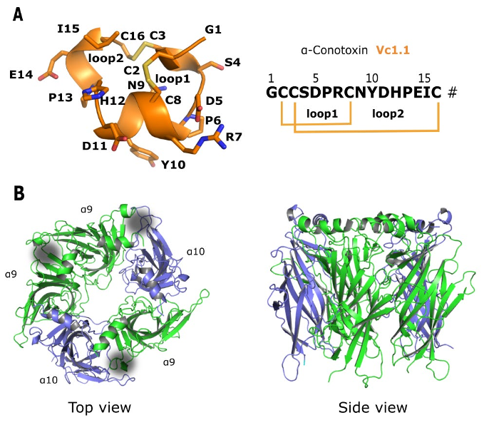
with a most prominent example being α-conotoxin Vc1.1 (Figure 1A).
13
Vc1.1 is a 16 amino acid, disulfide-bonded peptide identified from the venom of C.
victoria
14
and potently inhibits the α9α10 nAChR.
15-17
nAChRs are pentameric ligand-gated ion channels consisting of an extracellular
domain, a transmembrane domain and an intracellular domain and are expressed in
the central and peripheral nervous systems and non-neuronal cells.
18,19
The conotoxin
binding site is located at the extracellular domain contributed by the principal (+) and
complementary (−) components of two adjacent subunits (α1-α10, β1-β4, γ, δ or ε).
20
In the nervous system, they mediate the role of the neurotransmitter acetylcholine and
are involved in rapid synaptic transmission.
21-23
The non-neuronal functions of
nAChRs include cellular proliferation and regulation of the immune system. There are
many different nAChR subtypes with preferential distribution in the nervous system,

Citations
More filters
Journal ArticleDOI
TL;DR: The structure and function of the α9α10 nAChR are highlighted and studies of α-conotoxins targeting it are reviewed, including their three-dimensional structures, structure optimization strategies, and binding modes at the α 9α10nA chR, as well as their therapeutic potential.

22 citations

Journal ArticleDOI
06 Aug 2020-Toxins
TL;DR: This review scrutinises the N-terminal domain of the α-conotoxin family of peptides, a region defined by an invariant disulfide bridge, a turn-inducing proline residue and multiple polar sidechain residues, and focusses on structural features that provide analgesia through inhibition of high-voltage-activated Ca2+ channels.
Abstract: Several analgesic α-conotoxins have been isolated from marine cone snails. Structural modification of native peptides has provided potent and selective analogues for two of its known biological targets-nicotinic acetylcholine and γ-aminobutyric acid (GABA) G protein-coupled (GABAB) receptors. Both of these molecular targets are implicated in pain pathways. Despite their small size, an incomplete understanding of the structure-activity relationship of α-conotoxins at each of these targets has hampered the development of therapeutic leads. This review scrutinises the N-terminal domain of the α-conotoxin family of peptides, a region defined by an invariant disulfide bridge, a turn-inducing proline residue and multiple polar sidechain residues, and focusses on structural features that provide analgesia through inhibition of high-voltage-activated Ca2+ channels. Elucidating the bioactive conformation of this region of these peptides may hold the key to discovering potent drugs for the unmet management of debilitating chronic pain associated with a wide range of medical conditions.

20 citations

Journal ArticleDOI
TL;DR: A formerly defined rat α7 nAChR targeting α-CTx Mr1.1 was chemically synthesized and displayed analgesic activity in the rat chronic constriction injury (CCI) pain model and therefore presents a promising drug candidate.
Abstract: α-Conotoxins (α-CTxs) can selectively target nicotinic acetylcholine receptors (nAChRs) and are important drug leads for the treatment of cancer, chronic pain, and neuralgia. Here, we chemically synthesized a formerly defined rat α7 nAChR targeting α-CTx Mr1.1 and evaluated its activity at human nAChRs. Mr1.1 was most potent at the human (h) α9α10 nAChR with a half-maximal inhibitory concentration (IC50) of 92.0 nM. Molecular dynamic simulations suggested that Mr1.1 favorably binds at the α10(+)α9(-) and α9(+)α9(-) sites via hydrogen bonds and salt bridges, stabilizing the channel in a closed conformation. Although Mr1.1 and another antagonist, α-CTx Vc1.1 share high sequence similarity and disulfide-bond framework, Mr1.1 has distinct orientations at hα9α10. Based on the Mr1.1-hα9α10 model, analogues were generated, and the more potent Mr1.1[S4Dap], antagonized hα9α10 with an IC50 of 4.0 nM. Furthermore, Mr1.1[S4Dap] displayed analgesic activity in the rat chronic constriction injury (CCI) pain model and therefore presents a promising drug candidate.

7 citations

Proceedings ArticleDOI
17 Oct 2019
TL;DR: The topological landscape of the conopeptides were influenced by the Cα backbone and the nature of the intervening amino acid, and are predominantly electron-poor regions, allowing them to act as Lewis acids, and may play a role in their ability to interact with ACh receptors.
Abstract: Conopeptides are small, disulfide-rich polypeptides that have great potential as sources of possible drug candidates due to their activity against membrane receptors and ion channels. A challenge to the faster high-throughput in silico screening of these potential drug candidates is their diversity in structure and relatively low sequence similarity despite similar functions. In this study, the conopeptides of the α-pharmacological family is studied based on their Cα backbone, surface topology and sequence analysis. Structural alignment using FATCAT shows good alignment of the conopeptides based on their RMSD values. The main factor contributing to the homology of their structures is not only the Cys (Cys) framework forming the disulfide bridges but also the number of intervening amino acids between the Cys residues and the length of the polypeptide. The topological landscape of the conopeptides were influenced by the Cα backbone and the nature of the intervening amino acid, and are predominantly electron-poor regions, allowing them to act as Lewis acids. This may play a role in their ability to interact with ACh receptors.

5 citations


Cites background from "α-Conotoxin Vc1.1 Structure-Activit..."

  • ...Indeed, a large number of subclassifications of receptors have been discovered using conopeptide probes [6], [10], [11], [12], [13]....

    [...]

Journal ArticleDOI
TL;DR: In this review, the purpose of the review is to briefly show what different compounds of marine origin, from low molecular weight ones to peptides and proteins, offer for understanding the structure and mechanism of action of nicotinic acetylcholine receptors (nAChRs) and for finding novel drugs to combat the diseases where nA ChRs may be involved.
Abstract: The purpose of our review is to briefly show what different compounds of marine origin, from low molecular weight ones to peptides and proteins, offer for understanding the structure and mechanism of action of nicotinic acetylcholine receptors (nAChRs) and for finding novel drugs to combat the diseases where nAChRs may be involved. The importance of the mentioned classes of ligands has changed with time; a protein from the marine snake venom was the first excellent tool to characterize the muscle-type nAChRs from the electric ray, while at present, muscle and α7 receptors are labeled with the radioactive or fluorescent derivatives prepared from α-bungarotoxin isolated from the many-banded krait. The most sophisticated instruments to distinguish muscle from neuronal nAChRs, and especially distinct subtypes within the latter, are α-conotoxins. Such information is crucial for fundamental studies on the nAChR revealing the properties of their orthosteric and allosteric binding sites and mechanisms of the channel opening and closure. Similar data are provided by low-molecular weight compounds of marine origin, but here the main purpose is drug design. In our review we tried to show what has been obtained in the last decade when the listed classes of compounds were used in the nAChR research, applying computer modeling, synthetic analogues and receptor mutants, X-ray and electron-microscopy analyses of complexes with the nAChRs, and their models which are acetylcholine-binding proteins and heterologously-expressed ligand-binding domains.

5 citations

References
More filters
Journal ArticleDOI
TL;DR: The binding modes of α-conotoxin ImI to the α7-nAChR, currently the best-studied system experimentally, were investigated using comparative modeling and molecular dynamics simulations and the methodological developments reported here open avenues for computational scanning studies of a rapidly expanding range of wild-type and chemically modified α- Conotoxins.
Abstract: α-Conotoxins potently inhibit isoforms of nicotinic acetylcholine receptors (nAChRs), which are essential for neuronal and neuromuscular transmission. They are also used as neurochemical tools to study nAChR physiology and are being evaluated as drug leads to treat various neuronal disorders. A number of experimental studies have been performed to investigate the structure-activity relationships of conotoxin/nAChR complexes. However, the structural determinants of their binding interactions are still ambiguous in the absence of experimental structures of conotoxin-receptor complexes. In this study, the binding modes of α-conotoxin ImI to the α7-nAChR, currently the best-studied system experimentally, were investigated using comparative modeling and molecular dynamics simulations. The structures of more than 30 single point mutants of either the conotoxin or the receptor were modeled and analyzed. The models were used to explain qualitatively the change of affinities measured experimentally, including some nAChR positions located outside the binding site. Mutational energies were calculated using different methods that combine a conformational refinement procedure (minimization with a distance dependent dielectric constant or explicit water, or molecular dynamics using five restraint strategies) and a binding energy function (MM-GB/SA or MM-PB/SA). The protocol using explicit water energy minimization and MM-GB/SA gave the best correlations with experimental binding affinities, with an R2 value of 0.74. The van der Waals and non-polar desolvation components were found to be the main driving force for binding of the conotoxin to the nAChR. The electrostatic component was responsible for the selectivity of the various ImI mutants. Overall, this study provides novel insights into the binding mechanism of α-conotoxins to nAChRs and the methodological developments reported here open avenues for computational scanning studies of a rapidly expanding range of wild-type and chemically modified α-conotoxins.

74 citations

Journal ArticleDOI
15 Mar 2012-Toxicon
TL;DR: The principles of conotoxins cyclization are illustrated with applications to the α- and χ- conotoxin classes, which have been implicated as leads for the treatment of pain and a range of other disorders including neuroprotection, schizophrenia, depression and cancer.

67 citations

Journal ArticleDOI
TL;DR: The potential use of alpha9alpha10 selective drugs in conditions such as noise-induced hearing loss, tinnitus and auditory processing disorders is discussed.

66 citations

Journal ArticleDOI
TL;DR: This minireview focuses on the interaction of alpha-conotoxins with neuronal nicotinic receptors in light of the new understanding of the receptor structure and builds homology models of the binding domain of any nAChR subtype and fit in different ligands using docking programs.
Abstract: Recent and increasing use of computational tools in the field of nicotinic receptors has led to the publication of several models of ligand-receptor interactions. These models are all based on the crystal structure at 2.7 Angstrom resolution of a protein related to the extracellular N-terminus of nicotinic acetylcholine receptors (nAChRs), the acetylcholine binding protein. In the absence of any X-ray or NMR information on nAChRs, this new structure has provided a reliable alternative to study the nAChR structure. We are now able to build homology models of the binding domain of any nAChR subtype and fit in different ligands using docking programs. This strategy has already been performed successfully for the docking of several nAChR agonists and antagonists. This minireview focuses on the interaction of alpha-conotoxins with neuronal nicotinic receptors in light of our new understanding of the receptor structure. Computational tools are expected to reveal the molecular recognition mechanisms that govern the interaction between alpha-conotoxins and neuronal nAChRs at the molecular level. An accurate determination of their binding modes on the neuronal nAChR may allow the rational design of alpha-conotoxin-based ligands with novel nAChR selectivity.

49 citations

Journal ArticleDOI
28 Sep 2015-Toxins
TL;DR: A more comprehensive characterisation of the role of α9α10-nAChRs in pain is crucial for understanding the analgesic action of conotoxins and for improved drug design.
Abstract: The α9α10-nicotinic acetylcholine receptor (nAChR) has been implicated in pain and has been proposed to be a novel target for analgesics. However, the evidence to support the involvement of the α9α10-nAChR in pain is conflicted. This receptor was first implicated in pain with the characterisation of conotoxin Vc1.1, which is highly selective for α9α10-nAChRs and is an efficacious analgesic in chronic pain models with restorative capacities and no reported side effects. Numerous other analgesic conotoxin and non-conotoxin molecules have been subsequently characterised that also inhibit α9α10-nAChRs. However, there is evidence that α9α10-nAChR inhibition is neither necessary nor sufficient for analgesia. α9α10-nAChR-inhibiting analogues of Vc1.1 have no analgesic effects. Genetically-modified α9-nAChR knockout mice have a phenotype that is markedly different from the analgesic profile of Vc1.1 and similar conotoxins, suggesting that the conotoxin effects are largely independent of α9α10-nAChRs. Furthermore, an alternative mechanism of analgesia by Vc1.1 and other similar conotoxins involving non-canonical coupling of GABAB receptors to voltage-gated calcium channels is known. Additional incongruities regarding α9α10-nAChRs in analgesia are discussed. A more comprehensive characterisation of the role of α9α10-nAChRs in pain is crucial for understanding the analgesic action of conotoxins and for improved drug design.

39 citations

Related Papers (5)
Frequently Asked Questions (1)
Q1. What are the contributions in "Α-conotoxin vc1.1 structure-activity relationship at the human α9α10 nicotinic acetylcholine receptor investigated by minimal side chain replacement" ?

In this paper, the authors proposed the α10 ( + ) -α9 ( − ) interface as the favorable binding site of Vc1.1 at the ( α9 ) 2 ( α10 ) 3 nAChR.