scispace - formally typeset
Search or ask a question
Journal ArticleDOI

A refined pharmacophore identifies potent 4-amino-7-chloroquinoline-based inhibitors of the botulinum neurotoxin serotype A metalloprotease.

TL;DR: Four new, potent inhibitors of a structurally diverse small molecule (non-peptidic) inhibitors of the botulinum neurotoxin serotype A (BoNT/A) light chain (LC) are identified, supporting the structure-based pharmacophore contribution to inhibition.
Abstract: We previously identified structurally diverse small molecule (non-peptidic) inhibitors (SMNPIs) of the botulinum neurotoxin serotype A (BoNT/A) light chain (LC). Of these, several (including antimalarial drugs) contained a 4-amino-7-chloroquinoline (ACQ) substructure and a separate positive ionizable amine component. The same antimalarials have also been found to interfere with BoNT/A translocation into neurons, via pH elevation of the toxin-mediated endosome. Thus, this structural class of small molecules may serve as dual-function BoNT/A inhibitors. In this study, we used a refined pharmacophore for BoNT/A LC inhibition to identify four new, potent inhibitors of this structural class (IC50's ranged from 3.2 to 17 μM). Molecular docking indicated that the binding modes for the new SMNPIs are consistent with those of other inhibitors that we have identified, further supporting our structure-based pharmacophore. Finally, structural motifs of the new SMNPIs, as well as two structure-based derivatives, were ...

Summary (2 min read)

Introduction

  • 1,2 As a result, these enzymes, which are responsible for the paralysis associated with botulism, are listed as category A (highest priority) biothreat agents by the Centers for Disease Control and Prevention (CDC).
  • 2-6 Furthermore, as BoNTs are now used to treat a range of medical conditions, and in many cosmetic applications,3,7-14 they are being produced in increasing quantities, making their misuse, accidental overdosing, and/or instances of adverse side effects15 more likely.
  • The authors build on their previous work by describing how the refined pharmacophore33 was used to discover new small molecule inhibitors possessing the ACQ substructure and a separate ionizable, aliphatic amine component.

Results and Discussion

  • Based on the identification ofN,N-bis(7-chloroquinolin-4yl)diamines and five antimalarial drugs (amodiaquine, chloroquine, quinacrine, quinidine, and quinine) as SMNPIs of the BoNT/A LC,31 the authors have continued to pursue the identification of new inhibitors of this structural class possessing the weakly basic ACQ substructure and an ionizable amine; both of these components have been found to be key to activity.
  • Å from the centroid of the ACQ ), and (3) at least one of the new components from their expanded pharmacophore, either F33 (a positive ionizable moiety located 11.7-16.7 Å from the quinoline centroid) or G33 (a hydrophobic moiety located 8.5-12.5 Å from the quinoline centroid) .
  • Compounds1-3 were initially examined for percent inhibition of the BoNT/A LC at 50µM concentrations, while4 was tested at a 20µM concentration.
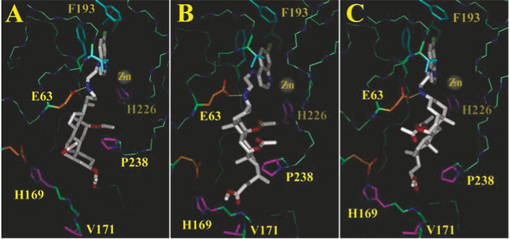
  • Molecular Docking of SMNPIs 1-4 Demonstrates a Consistency with Previous Inhibitor Binding Modes, Reinforcing The authors Structure-Based Pharmacophore Approach.
  • Substituents of1-3 and one of the ACQs of4 engage in favorable hydrophobic contacts with residues Phe 162, Phe 163, and Thr 219 of hydrophobic binding subsite 131-33 (also described as the S1′ binding site34).

Component E Dominates Potency for This Structural Class.

  • The final analyses of this study involved determining the importance of the structural components of1-4 to BoNT/A LC inhibition.
  • To determine if incorporating pharmacophore component E might transform8 into an inhibitor, the 3R-acetyloxy group of this molecule was replaced with an ionizable 3R-NH2 substituent , effectively creating a substructure of SMNPI3 (Scheme 1), the most potent molecule of the ACQ-cholate acetate congener series (Table 1).
  • Finally, to determine if it would be possible to restore the inhibitory potency of10, the 3R-O-cholate-acetate and ACQ components were tethered with an extended linker incorporating a central, secondary amine.
  • Specifically, the extended linker forces the cholate-acetate portion of11 to adopt a shallower binding mode, with its C23 methoxycarbonyl oriented outside of the substrate binding cleft .
  • Amino ether11 (43% from 14) was obtained in a reductive amination reaction between 6 and aldehyde15 (Scheme 1).

Conclusion

  • An expanded/refined pharmacophore for BoNT/A LC inhibition33 was used to identify four new, potent SMNPIs of the ACQ structural class.
  • This provides additional support that molecular scaffolds, which are larger than many reported BoNT/A LC inhibitors,31,40-42 are needed to provide superior inhibition.
  • Finally, examination of substructures composing1-4 (i.e., compounds5-9), as well as derivatives 10 and 11, revealed the critical importance of positive-ionizable pharmacophore component E to the potencies of inhibitors in the structural class.

Did you find this useful? Give us your feedback

Content maybe subject to copyright    Report

%4/<+89/:?5,+(8'91'/4)524%4/<+89/:?5,+(8'91'/4)524
/-/:'2533549%4/<+89/:?5,+(8'91'/4)524/-/:'2533549%4/<+89/:?5,+(8'91'/4)524
%#83?"+9+'8). %#+6'8:3+4:5,+,+49+

"+A4+*!.'83')56.58+*+4:/A+9!5:+4:"+A4+*!.'83')56.58+*+4:/A+9!5:+4:
3/45).25857;/452/4+'9+*4./(/:5895,:.+5:;2/4;33/45).25857;/452/4+'9+*4./(/:5895,:.+5:;2/4;3
+;85:5>/4#+85:?6++:'225685:+'9++;85:5>/4#+85:?6++:'225685:+'9+
'3+9;84+::
+0'4 69+4/)'
'3'8'0#8/8'-.'<'4
"+1.'!'4).'2
58*54";:.+2
#++4+>:6'-+,58'**/:/54'2';:.589
5225=:./9'4*'**/:/54'2=5819':.::69*/-/:'2)533549;42+*;;9'83?8+9+'8).
!'8:5,:.+ 6+8':/549"+9+'8).#?9:+394-/4++8/4-'4*4*;9:8/'24-/4++8/4-533549
;84+::'3+9 69+4/)'+0'4#8/8'-.'<'4'3'8'0!'4).'2"+1.'";:.+258*54+8354+
44"-;?+4$'3+44?$'8''4+5;-2'9)8':.54458#).3/*:'3+9
&+44+89:85354':.'4;99/5"/)1#52'0'5-*'4'4*'<'8/#/4'"+A4+*!.'83')56.58+
*+4:/A+9!5:+4:3/45).25857;/452/4+'9+*4./(/:5895,:.+5:;2/4;3+;85:5>/4#+85:?6+
+:'225685:+'9+
%#83?"+9+'8).

.::69*/-/:'2)533549;42+*;;9'83?8+9+'8).
$./98:/)2+/9(85;-.::5?5;,58,8++'4*56+4'))+99(?:.+%#+6'8:3+4:5,+,+49+':
/-/:'2533549%4/<+89/:?5,+(8'91'/4)524:.'9(++4'))+6:+*,58/4)2;9/54/4%#83?"+9+'8).(?'4
';:.58/@+*'*3/4/9:8':585,/-/:'2533549%4/<+89/:?5,+(8'91'/4)524

;:.589;:.589
'3+9;84+::+0'4 69+4/)''3'8'0#8/8'-.'<'4"+1.'!'4).'258*54";:.+244"
+8354+$'3-;?+4$'8'+44?5;-2'9'4+54458)8':.'3+9#).3/*:
54':.'4&+44+89:853"/)1;99/55-*'4#52'0''4*#/4''<'8/
$./9'8:/)2+/9'<'/2'(2+':/-/:'2533549%4/<+89/:?5,+(8'91'/4)524.::69*/-/:'2)533549;42+*;
;9'83?8+9+'8).

A Refined Pharmacophore Identifies Potent 4-Amino-7-chloroquinoline-Based Inhibitors of the
Botulinum Neurotoxin Serotype A Metalloprotease
James C. Burnett,
Dejan Opsenica,
Kamaraj Sriraghavan,
§
Rekha G. Panchal,
Gordon Ruthel,
|
Ann R. Hermone,
Tam L. Nguyen,
Tara A. Kenny,
Douglas J. Lane,
Connor F. McGrath,
James J. Schmidt,
|
Jonathan L. Vennerstrom,
§
Rick Gussio,
Bogdan A. Sˇolaja,*
,#
and Sina Bavari*
,|
SAIC-Frederick, Inc., Target Structure-Based Drug DiscoVery Group, Frederick, Frederick, Inc., National Cancer Institute at Frederick,
P.O. Box B, F.V.C. 310, Frederick, Maryland 21702, The Institute of Chemistry, Technology, and Metallurgy, NjegosˇeVa 12, YU-11001
Belgrade, Serbia, College of Pharmacy, 986025 UniVersity of Nebraska Medical Center, Omaha, Nebraska 68198, U.S. Army Medical
Research Institute of Infectious Diseases, Fort Detrick, 1425 Porter Street, Frederick, Maryland 21702, DeVelopmental Therapeutics Program,
P.O. Box B, F.V.C. 310, NCI Frederick, Frederick, Maryland 21702, and Faculty of Chemistry, The UniVersity of Belgrade, Studentski trg 16,
P.O. Box 158, YU-11001 Belgrade, Serbia
ReceiVed December 19, 2006
We previously identified structurally diverse small molecule (non-peptidic) inhibitors (SMNPIs) of the
botulinum neurotoxin serotype A (BoNT/A) light chain (LC). Of these, several (including antimalarial drugs)
contained a 4-amino-7-chloroquinoline (ACQ) substructure and a separate positive ionizable amine component.
The same antimalarials have also been found to interfere with BoNT/A translocation into neurons, via pH
elevation of the toxin-mediated endosome. Thus, this structural class of small molecules may serve as dual-
function BoNT/A inhibitors. In this study, we used a refined pharmacophore for BoNT/A LC inhibition to
identify four new, potent inhibitors of this structural class (IC
50
’s ranged from 3.2 to 17 µM). Molecular
docking indicated that the binding modes for the new SMNPIs are consistent with those of other inhibitors
that we have identified, further supporting our structure-based pharmacophore. Finally, structural motifs of
the new SMNPIs, as well as two structure-based derivatives, were examined for activity, providing valuable
information about pharmacophore component contributions to inhibition.
Introduction
Botulinum neurotoxins (BoNTs)
a
are the most potent of the
biological toxins; the lethal dose of BoNT serotype A (BoNT/
A) is estimated to be between 1 and 5 ng kg
-1
for humans.
1,2
As a result, these enzymes, which are responsible for the
paralysis associated with botulism, are listed as category A
(highest priority) biothreat agents by the Centers for Disease
Control and Prevention (CDC). BoNTs are easily produced and
may be delivered via food “spiking” and/or aerosol route.
2-6
Furthermore, as BoNTs are now used to treat a range of medical
conditions, and in many cosmetic applications,
3,7-14
they are
being produced in increasing quantities, making their misuse,
accidental overdosing, and/or instances of adverse side effects
15
more likely. Neither the currently available CDC BoNT equine
antitoxins, which can cause adverse anaphylaxis and serum
sickness,
16
nor experimental antibodies can counter these
enzymes once they are inside neurons. Furthermore, BoNT
intoxication can occur rapidly,
17
and individuals who have been
maliciously exposed to a BoNT(s), or have received an
accidental overdose, will most likely seek medical attention only
after clinical symptoms (i.e., muscle paralysis) manifest. At this
time, critical care mechanical ventilation is the only life-saving
option once diaphragm muscles cease to function. Yet, the
effects of internalized BoNTs can last for weeks,
18,19
rendering
such medical care impractical for wide scale application. By
comparison, small molecule (non-peptidic) inhibitors (SMNPIs)
could serve as post-intoxication “rescue” therapeutics and
prophylactics.
There are seven known BoNT serotypes (identified as A-F).
Each cleaves a component of the SNARE (soluble N-ethylma-
leimide-sensitive factor attachment protein receptor) com-
plex,
20,21
which facilitates the transport of acetylcholine into
neuromuscular junctions. BoNT serotypes A and E cleave
SNAP-25 (synaptosomal-associated protein (25 kDa)),
22
sero-
types B, D, F,
23
and G cleave VAMP (vesicle-associated
membrane protein),
24-27
and serotype C1 cleaves both SNAP-
25 and syntaxin 1.
28
X-ray crystal structures of BoNT holotoxins
29,30
show that
these enzymes are composed of a heavy chain (HC) and a light
chain (LC), which, following post-translational modification,
are connected by a disulfide bridge.
29
The LC is a zinc
metalloprotease. The HC transports the LC into the neuronal
cytosol via an acidic endosome.
In a previous publication,
31
we identified a range of novel,
structurally diverse, non-zinc chelating SMNPIs of the BoNT/A
LC. In particular, we showed that several compounds containing
the weakly basic (pK
a
8) ACQ substructure and a separate
ionizable amine component were among the most potent of the
inhibitors.
31
Additionally, we proposed binding modes for the
compounds and developed a common pharmacophore for
BoNT/A LC inhibition.
31
In a subsequent molecular dynamics
study, we demonstrated that conformationally flexible loops
surrounding the BoNT/A LC substrate binding cleft may reorient
to decrease the solvent accessibility of the cleft (as compared
to respective energy refined structures), while simultaneously
providing more hydropathically feasible binding contacts for
* To whom correspondence should be addressed. (B.A.S.) Phone: (+381-
11) 638-606. Fax: (+381-11) 638-061. E-mail: bsolaja@chem.bg.ac.yu.
(S.B.) Phone: (301) 619-4246. Fax: (301) 619-2348. E-mail:
sina.bavari@us.army.mil.
National Cancer Institute at Frederick.
The Institute of Chemistry, Technology, and Metallurgy.
§
University of Nebraska Medical Center.
|
U.S. Army Medical Research Institute of Infectious Diseases.
Developmental Therapeutics Program.
#
The University of Belgrade.
a
Abbreviations: BoNTs, botulinum neurotoxins; BoNT/A, botulinum
neurotoxin serotype A; BoNT/A LC, BoNT/A light chain; SMNPIs, small
molecule (non-peptidic) inhibitors; ACQ, 4-amino-7-chloroquinoline.
2127J. Med. Chem. 2007, 50, 2127-2136
10.1021/jm061446e CCC: $37.00 © 2007 American Chemical Society
Published on Web 04/07/2007
This article is a U.S. government work, and is not subject to copyright in the United States.

SMNPIs.
32
The molecular dynamics studies were pivotal for
identifying a binding mode
33
for the potent pseudo-peptide
inhibitor 2-mercapto-3-phenylpropionyl-RATKML (mpp-RAT-
KML, K
i
) 330 nM
34
). In the same study, we also proposed
new pharmacophore components and constraints based on the
docked model of mpp-RATKML, and, using this information,
we identified more potent SMNPIs.
33
In this study, we build
on our previous work by describing how the refined pharma-
cophore
33
was used to discover new small molecule inhibitors
possessing the ACQ substructure and a separate ionizable,
aliphatic amine component.
Results and Discussion
Based on the identification of N,N-bis(7-chloroquinolin-4-
yl)diamines and five antimalarial drugs (amodiaquine, chloro-
quine, quinacrine, quinidine, and quinine) as SMNPIs of the
BoNT/A LC,
31
we have continued to pursue the identification
of new inhibitors of this structural class possessing the weakly
basic ACQ substructure and an ionizable amine; both of these
components have been found to be key to activity.
31
Further
supporting this approach are the findings of Deshpande et al.
35
and Sheridan et al.,
36
demonstrating that ACQ-based drugs,
which also possess a separate ionizable nitrogen atom (e.g.,
amodiaquine), delay BoNT/A induced muscle paralysis. In those
studies, it was hypothesized that the compounds act by interfer-
ing with toxin translocation, most likely by increasing the pH
of the toxin-mediated endosome.
35,36
This is a logical supposition
as ACQ antimalarials have been shown to target and increase
the pH of the acidic food vacuole of Plasmodium falciparum.
37-39
However, neither of the earlier studies reported LC inhibition
in vitro. As indicated above, we demonstrated that several of
the same antimalarials also directly inhibit BoNT/A LC met-
alloprotease activity;
31
however, they do so at higher concentra-
tions than reported in the muscle twitch assays used in the earlier
studies.
35,36
This comparison indicates that the observed muscle
twitch inhibition could not have resulted solely from inhibition
of the enzyme’s LC. Thus, the Deshpande et al.
35
and Sheridan
et al.
36
data, in combination with our previous findings, indicate
that such molecules may serve as dual-function countermeasures,
acting both as translocation inhibitors and directly against the
metalloprotease activity of the LC.
A Refined Pharmacophore Is Used To Identify New
BoNT/A LC Inhibitors Possessing Both the ACQ Substruc-
ture and a Separate Ionizable Amine. The first step in
identifying new SMNPIs was to use a recently refined/expanded
pharmacophore for BoNT/A LC inhibition
33
(Figure 1A) to
evaluate diverse target compounds possessing the ACQ sub-
structure. It is important to note that the eight-component
pharmacophore model presented in Figure 1A is a logical
extension of the six-component model shown in our original
work
31
and is part of an ongoing refinement of our pharma-
cophore for BoNT/A LC inhibition
32,33
(which is occurring as
more data become available). In this study, we wanted to identify
compounds containing as many pharmacophore features as
possible, but with the following specific components: (1) the
ACQ substructure (which incorporates pharmacophore compo-
nents A, a heteroatom associated with A, and C (Figure 1A)),
(2) an ionizable amine located 6.5-9.5 Å from the centroid of
the ACQ (pharmacophore component E (Figure 1A)), and (3)
at least one of the new components from our expanded
pharmacophore, either F
33
(a positive ionizable moiety located
11.7-16.7 Å from the quinoline centroid) or G
33
(a hydrophobic
moiety located 8.5-12.5 Å from the quinoline centroid) (Figure
1A). Subsequently, a congeneric series of 4-amino-7-chloro-
quinoline-cholate-acetates, 1-3, and a tri-ACQ derivative, 4,
were identified (see Scheme 1 for two-dimensional (2-D)
structures of 1-3 and Figure 2 for the 2-D structure of 4).
Pharmacophore mapping to 1-4 indicated that in each case
ACQ is plane A (Figure 1B-E), and the quinoline ring nitrogen
is the heteroatom associated with this plane. For 1-3, compo-
nent B is the central decahydronapthalene of the methyl cholate
acetate (rings B and C according to steroid ring designation)
(Figure 1B-D), while in 4 it is a second ACQ (Figure 1E).
Hydrophobic component C is the 7-chloro substituent of the
plane A ACQ for all inhibitors (Figure 1B-E), and pharma-
cophore component D is a methyl for 1 (Figure 1B), an acetyl
group for 2 and 3 (Figure 1C and D), and a chloro substituent
for 4 (Figure 1E).
As a second specific criterion, each of the molecules contains
a separate ionizable, aliphatic amine (Figure 1B-E), which
corresponds to pharmacophore component E (Figure 1A).
Finally, all of the molecules possess hydrophobic pharmacoph-
ore component G from the expanded/refined pharmacophore
33
(Figure 1A) (the third specified criterion, see above), which is
an acetyl group for 1 and 2 (Figure 1B and C), a methyl group
for 3 (Figure 1D), and a chloro substituent for 4 (Figure 1E).
Figure 1. Pharmacophore mapping to inhibitors 1-4. (A) The refined/
expanded pharmacophore for BoNT/A LC inhibition. Planar compo-
nents A and B are magenta rectangles. The dashed, green circle in plane
A represents a heteroatom. Hydrophobic components C and D are
shown as dashed, light blue circles. The positive ionizable pharma-
cophore component E is shown as a dashed, red circle. New pharma-
cophore component F,
33
also a positive ionizable moiety, is shown as
a dashed red circle, while new hydrophobic pharmacophore component
G is a dashed orange circle. (B, C, D, and E) Mapping of inhibitors
1-4, respectively, to the expanded pharmacophore. Distances between
components are in angstroms. Pharmacophore component colors are
as indicated in (A).
2128 Journal of Medicinal Chemistry, 2007, Vol. 50, No. 9 Burnett et al.

As observed in Figure 1B-E, 1-4 all show a good fit with
respect to the distance constraints specified by the pharmacoph-
ore.
In Vitro Testing. Compounds 1-3 were initially examined
for percent inhibition of the BoNT/A LC at 50 µM concentra-
tions, while 4 was tested at a 20 µM concentration. Results from
these initial analyses indicated 90% enzyme inhibition by 1 and
3, 80% inhibition by 2, and 67% inhibition by 4. Subsequent
determination of IC
50
values revealed that SMNPIs 1-4 are
among the most potent inhibitors of the BoNT/A LC reported
to date,
33,40-42
possessing IC
50
values ranging from 3.2 to 17.0
µM (Table 1). In agreement with structurally distinct SMNPIs
reported by our research group (possessing K
i
values ranging
from 3.0 to 10.0 µM
33
), compounds 1-4 demonstrate that our
strategy of incorporating new pharmacophore features (i.e.,
pharmacophore components F or G),
33
in addition to original
components,
31,32
results in the identification of more potent
inhibitors.
Molecular Docking of SMNPIs 1-4 Demonstrates a
Consistency with Previous Inhibitor Binding Modes, Rein-
forcing Our Structure-Based Pharmacophore Approach.
Compounds 1-4 were docked in the BoNT/A LC substrate
binding cleft to determine if they would also engage in
intermolecular contacts that were comparable to those predicted
for other structurally diverse SMNPIs.
31-33
Similar to our
previously docked models of ACQ inhibitors,
31,32
the 7-chloro
Scheme 1
a
a
Reagents and conditions: (a) (i) MsCl, pyridine; (ii) 6, DMF; (b) 6, NaBH
3
CN, CH
3
CN; (c) allyl bromide,
i
Pr
2
NEt, NMP, 80 °C; (d) (i) O
3
, MeOH, -78
°C; (ii) Me
2
S; (e) 6, NaBH(OAc)
3
,CH
2
Cl
2
; (f) NaBH(OAc)
3
,CH
2
Cl
2
; (g) (i) MsCl, pyridine; (ii) NaN
3
, DMF; (h) PPh
3
, THF, H
2
O, 50 °C; (i) 4,7-
dichloroquinoline, PhOH, 120 °C.
Figure 2. Two-dimensional structures of 4-9.
Table 1. Inhibitory Potencies of 1-4
SMNPI IC
50
a
(µM)
1 10 ((0.80)
2 17 ((1.7)
3 7.0 ((1.0)
4 3.2 ((0.92)
a
IC
50
values for the SMNPIs were calculated from plots of concentration
versus inhibition (see Experimental Section for details). Results are the
averages of duplicate determinations.
Pharmacophore Identifies Inhibitors of Botulinum A Journal of Medicinal Chemistry, 2007, Vol. 50, No. 9 2129

Citations
More filters
Journal ArticleDOI
01 Nov 2012-Toxicon
TL;DR: The findings show the value of VS in identifying potential inhibitors of the toxin for further development and improvement and the importance of favorable energetic and glide criteria.

7 citations

Journal ArticleDOI
TL;DR: In this article, the structural and spectroscopic characteristics of the synthesized structurally novel compound 4-chloro-6-methylquinoline-2(1H)-one (4C6MQ) and its isomer 4C8MQ have been examined by means of experimental and computational quantum chemical methods like density functional theory (DFT).
Abstract: The structural and spectroscopic characteristics of the synthesized structurally novel compound 4-chloro-6-methylquinoline-2(1H)-one (4C6MQ) and its isomer 4-chloro-8-methylquinoline-2(1H)-one (4C8MQ) have been examined by means of experimental and computational quantum chemical methods like density functional theory (DFT). The crystal structure of the 4C6MQ compound has been brought to light by single-crystal x-ray diffraction (SCXRD) method which consists of two independent molecules (A and B) in the asymmetric unit with similar conformations. Both the isomer compounds are characterized spectroscopically by FTIR, FT-Raman, UV-Vis, and NMR spectrum and compared with DFT results. The geometries of the isomer compounds have been optimized by using DFT/B3LYP method with the 6-311G++(d,p) basis sets. From the optimized geometry of the compounds, geometric parameters (bond lengths, bond angles, and torsion angles); vibrational analysis; chemical shifts; and electronic absorption of the isomer compounds have been computed and compared with the experimental result. The detailed assignments of vibrational wave numbers have been prepared based on potential energy distribution (PED) which was carried out in the VEDA4 program. In addition, natural bonding orbital analysis, frontier molecular orbital, and molecular electrostatic potential have been explained theoretically. The in silico (absorption, distribution, metabolism, excretion and toxicity) studies were analyzed to identify the potential drug likeliness of the isomer compounds. The implications of the inhibitory activity of isomer compounds against DNA gyrase and lanosterol 14 α-demethylase enzyme by molecular docking are discussed. Further, the isomer compounds were screened for their antibacterial and antifungal activities.

7 citations

Journal ArticleDOI
TL;DR: Clinical uses of BoNT continue to expand at an ever-increasing rate; however, further optimization ofBoNT therapy is critical to maximize efficacy, and is a valid target for the development of pharmacological interventions to prevent the considerable mortality associated with botulism.
Abstract: Botulinum neurotoxin (BoNT) has recently catapulted into the public consciousness in two seemingly disparate roles: potential use as a biological weapon and treatment of neuromuscular disorders. This bacterially produced protein is the most potent toxin known to humans, with a lethal dose estimated at approximately 1 ng/kg of body weight. BoNT intoxication occurs via a multistep process that includes recognition of peripheral neuronal cell membrane receptors, endocytosis and translocation of the light chain metalloprotease into the cytosol, and catalytic cleavage of soluble N-ethylmaleimide-sensitive factor attachment protein receptor (SNARE) proteins critical for vesicle fusion and neurotransmitter release. Each of these distinct steps has been studied at the molecular level and is a valid target for the development of pharmacological interventions to prevent the considerable mortality associated with botulism. Alternatively, clinical uses of BoNT continue to expand at an ever-increasing rate; however, f...

5 citations

Journal ArticleDOI
TL;DR: In this article, the interactions between proline and some aromatic moieties in ligands were investigated by means of the density functional theory using an exchange-correlation functional capable of taking into account dispersion interactions.
Abstract: Proline, due to its conformational specificity, is known to show some unique properties and has significant functions in the tertiary structure of proteins. It was suggested that proline could have an important influence on some vital interactions in protein as well, by engaging in non-covalent stabilization interactions with some aromatic moieties. In this work, the interactions that occur between proline and some aromatic moieties in ligands were investigated by means of the density functional theory using an exchange–correlation functional capable of taking into account dispersion interactions. The obtained results showed that the stabilization energy between a properly placed proline and an aromatic moiety could be as large as 25 kJ/mol and hence be a significant factor in placing a ligand in binding site of a protein. This indicates that the error in determining the most favorable structure of ligand–protein complexes obtained by usual molecular docking experiments sometimes could be the result of neglecting this type of interactions.

5 citations

Journal ArticleDOI
TL;DR: Steroidal derivative 16 appears to be the first example of LC inhibitors antagonizing BoNT intoxication in mouse embryonic stem cell derived motor neurons (mES-MNs) in a postexposure model, and the favorable in vitro ADMET results strongly support further work in this series.
Abstract: The synthesis and inhibitory potencies against botulinum neurotoxin serotype A light chain (BoNT/A LC) using in vitro HPLC based enzymatic assay for various steroidal, benzothiophene, thiophene, and adamantane 4-aminoquinoline derivatives are described In addition, the compounds were evaluated for the activity against BoNT/A holotoxin in mouse embryonic stem cell derived motor neurons Steroidal derivative 16 showed remarkable protection (up to 89% of uncleaved SNAP-25) even when administered 30 min postintoxication This appears to be the first example of LC inhibitors antagonizing BoNT intoxication in mouse embryonic stem cell derived motor neurons (mES-MNs) in a postexposure model Oral administration of 16 was well tolerated in the mouse up to 600 mg/kg, qd Although adequate unbound drug levels were not achieved at this dose, the favorable in vitro ADMET results strongly support further work in this series

5 citations

References
More filters
Journal ArticleDOI
29 Oct 1992-Nature
TL;DR: The results indicate that tetanus and botulinum B neurotoxins block neurotransmitter release by cleaving synaptobrevin-2, a protein that, on the basis of the results, seems to play a key part in neurotransmitterRelease.
Abstract: Clostridial neurotoxins, including tetanus toxin and the seven serotypes of botulinum toxin (A-G), are produced as single chains and cleaved to generate toxins with two chains joined by a single disulphide bond (Fig. 1). The heavy chain (M(r) 100,000 (100K)) is responsible for specific binding to neuronal cells and cell penetration of the light chain (50K), which blocks neurotransmitter release. Several lines of evidence have recently suggested that clostridial neurotoxins could be zinc endopeptidases. Here we show that tetanus and botulinum toxins serotype B are zinc endopeptidases, the activation of which requires reduction of the interchain disulphide bond. The protease activity is localized on the light chain and is specific for synaptobrevin, an integral membrane protein of small synaptic vesicles. The rat synaptobrevin-2 isoform is cleaved by both neurotoxins at the same single site, the peptide bond Gln 76-Phe 77, but the isoform synaptobrevin-1, which has a valine at the corresponding position, is not cleaved. The blocking of neurotransmitter release of Aplysia neurons injected with tetanus toxin or botulinum toxins serotype B is substantially delayed by peptides containing the synaptobrevin-2 cleavage site. These results indicate that tetanus and botulinum B neurotoxins block neurotransmitter release by cleaving synaptobrevin-2, a protein that, on the basis of our results, seems to play a key part in neurotransmitter release.

1,727 citations

Journal ArticleDOI
28 Feb 2001-JAMA
TL;DR: People potentially exposed to botulinum toxin should be closely observed, and those with signs of botulism require prompt treatment with antitoxin and supportive care that may include assisted ventilation for weeks or months.
Abstract: ObjectiveThe Working Group on Civilian Biodefense has developed consensus-based recommendations for measures to be taken by medical and public health professionals if botulinum toxin is used as a biological weapon against a civilian population.ParticipantsThe working group included 23 representatives from academic, government, and private institutions with expertise in public health, emergency management, and clinical medicine.EvidenceThe primary authors (S.S.A. and R.S.) searched OLDMEDLINE and MEDLINE (1960–March 1999) and their professional collections for literature concerning use of botulinum toxin as a bioweapon. The literature was reviewed, and opinions were sought from the working group and other experts on diagnosis and management of botulism. Additional MEDLINE searches were conducted through April 2000 during the review and revisions of the consensus statement.Consensus ProcessThe first draft of the working group's consensus statement was a synthesis of information obtained in the formal evidence-gathering process. The working group convened to review the first draft in May 1999. Working group members reviewed subsequent drafts and suggested additional revisions. The final statement incorporates all relevant evidence obtained in the literature search in conjunction with final consensus recommendations supported by all working group members.ConclusionsAn aerosolized or foodborne botulinum toxin weapon would cause acute symmetric, descending flaccid paralysis with prominent bulbar palsies such as diplopia, dysarthria, dysphonia, and dysphagia that would typically present 12 to 72 hours after exposure. Effective response to a deliberate release of botulinum toxin will depend on timely clinical diagnosis, case reporting, and epidemiological investigation. Persons potentially exposed to botulinum toxin should be closely observed, and those with signs of botulism require prompt treatment with antitoxin and supportive care that may include assisted ventilation for weeks or months. Treatment with antitoxin should not be delayed for microbiological testing.

1,659 citations

Journal ArticleDOI
TL;DR: The crystal structure of the entire 1,285 amino acid di-chain neurotoxin was determined and the toxin appears as a hybrid of varied structural motifs and suggests a modular assembly of functional subunits to yield pathogenesis.
Abstract: Botulinum neurotoxin type A (BoNT/A) is the potent disease agent in botulism, a potential biological weapon and an effective therapeutic drug for involuntary muscle disorders. The crystal structure of the entire 1,285 amino acid di-chain neurotoxin was determined at 3.3 A resolution. The structure reveals that the translocation domain contains a central pair of alpha-helices 105 A long and a approximately 50 residue loop or belt that wraps around the catalytic domain. This belt partially occludes a large channel leading to a buried, negative active site--a feature that calls for radically different inhibitor design strategies from those currently used. The fold of the translocation domain suggests a mechanism of pore formation different from other toxins. Lastly, the toxin appears as a hybrid of varied structural motifs and suggests a modular assembly of functional subunits to yield pathogenesis.

737 citations

Journal ArticleDOI
TL;DR: It is concluded that HPC‐1/syntaxin, a membrane protein present in axonal and synaptic membranes, is involved in exocytotic membrane fusion.
Abstract: The anaerobic bacterium Clostridium botulinum produces several related neurotoxins that block exocytosis of synaptic vesicles in nerve terminals and that are responsible for the clinical manifestations of botulism. Recently, it was reported that botulinum neurotoxin type B as well as tetanus toxin act as zinc-dependent proteases that specifically cleave synaptobrevin, a membrane protein of synaptic vesicles (Link et al., Biochem. Biophys. Res. Commun., 189, 1017-1023; Schiavo et al., Nature, 359, 832-835). Here we report that inhibition of neurotransmitter release by botulinum neurotoxin type C1 was associated with the proteolysis of HPC-1 (= syntaxin), a membrane protein present in axonal and synaptic membranes. Breakdown of HPC-1/syntaxin was selective since no other protein degradation was detectable. In vitro studies showed that the breakdown was due to a direct interaction between HPC-1/syntaxin and the toxin light chain which acts as a metallo-endoprotease. Toxin-induced cleavage resulted in the generation of a soluble fragment of HPC-1/syntaxin that is 2-4 kDa smaller than the native protein. When HPC-1/syntaxin was translated in vitro, cleavage occurred only when translation was performed in the presence of microsomes, although a full-length product was obtained in the absence of membranes. However, susceptibility to toxin cleavage was restored when the product of membrane-free translation was subsequently incorporated into artificial proteoliposomes. In addition, a translated form of HPC-1/syntaxin, which lacked the putative transmembrane domain at the C-terminus, was soluble and resistant to toxin action. We conclude that HPC-1/syntaxin is involved in exocytotic membrane fusion.(ABSTRACT TRUNCATED AT 250 WORDS)

541 citations

Journal ArticleDOI
TL;DR: The structures of BoNT/B and its complex with sialyllactose provide a detailed description of the active site and a model for interactions between the toxin and its cell surface receptor, and the latter may provide valuable information for recombinant vaccine development.
Abstract: Clostridium botulinum neurotoxins are among the most potent toxins to humans. The crystal structures of intact C. botulinum neurotoxin type B (BoNT/B) and its complex with sialyllactose, determined at 1. 8 and 2.6 A resolution, respectively, provide insight into its catalytic and binding sites. The position of the belt region in BoNT/B is different from that in BoNT/A; this observation presents interesting possibilities for designing specific inhibitors that could be used to block the activity of this neurotoxin. The structures of BoNT/B and its complex with sialyllactose provide a detailed description of the active site and a model for interactions between the toxin and its cell surface receptor. The latter may provide valuable information for recombinant vaccine development.

450 citations

Related Papers (5)