Adaptive spatial compounding for improving ultrasound images of the epidural space on human subjects
Summary (4 min read)
1.1 Epidural anesthesia in obstetrics
- Anesthesia is injected into the epidural space as shown on Fig. 1 and a nerve block for the lower body is then provided.
- The catheter is then inserted through the epidural needle shaft into the epidural space.
- The ultrasound group achieved a success rate of 84% in the first 10 attempts whereas the control group had a success rate of 60%.
- Which is around 7mm and much smaller than the epidural depth of adults (20-90mm), it shows the potential benefit of using ultrasound to visualize the lumbar region during an epidural needle insertion procedure.
1.2 Ultrasound imaging of lumbar region
- Ultrasound is noninvasive, harmless at low power, portable, accurate and cost effective.
- Ultrasound of the lumbar region shows an image filled with speckle and artifacts which can impede detection of important features such as the ligamentum flavum and other hard to detect structures.
- The received echo is based on ultrasound reflections from large-scale (relative to wavelength) structures, such as bone (i.e. specular reflection) and reflections from small-scale structures, such as cells (i.e. random scattering).
- If the specular reflection is strong enough, such as a bone, it casts shadows in the beam direction and structures underneath are obstructed.
- Terms of Use: http://spiedl.org/terms with respect to the reflectors.
1.3 Image processing techniques
- Many post-processing methods employ filters to reduce speckle.
- Examples are the diffusion filter 10 , the adaptive weighted median filter11 , homogeneous region growing mean filter, and aggressive region growing filter 12 .
- Frequency compounding captures several images at the same location at different transmission frequencies, decorrelating the speckle patterns among images.
- Spatial compounding is now popular among commercial manufacturers and will be presented in the next section.
2.1 Spatial compounding
- Spatial compounding uses beam steering19 20 21 , which captures several frames by sending the ultrasound pulses at different angles of incidence (see Fig. 2(b) for an illustration of the principle).
- The application of spatial compounding to these images reduced speckle noise and improved the boundary continuity.
- Spatial compounding also has other benefits, such as the possibility of enhancing structures that are only visible at certain beam angles.
- Certain weak but important features, such as a biopsy needle 9 24 only appear at certain Proc. of SPIE Vol.
- The structure of main interest in this work is the epidural space, which is immediately under the ligamentum flavum and initial experience suggests it is only clear at certain beam angles.
2.2 Registration techniques
- Conventional spatial compounding still suffers from blurring due to misalignment of features.
- The speed of sound varies by as much as 14% in soft tissue25 and the resulting distortion (including refraction) causes the apparent positions of structures to be slightly different under different angles of incidence.
- Re-alignment of the features using an additional block-matching non-rigid registration was previously proposed by their group to properly align the structures of each image 13 .
- The result was a sharper ultrasound image.
- Building on those results, the warping/compounding method is extended here to improve visibility of the ligamentum flavum in vivo, and therefore the epidural space.
2.2.1 Similarity measures
- Registration performance is highly dependent on the similarity measure used as a cost function for finding the best alignment.
- A good similarity measure will yield a single strong peak upon best alignment.
- Previous literature used several methods such as sum of absolute differences (SAD), mean squared error (MSE) 27 , normalized covariance (NCOV), normalized crosscorrelation (NCC), entropy of the difference image and mutual information28 .
- Mutual information29 30 is a very popular similarity measure for registration of multimodality images but is too easily affected by artifacts.
- Since the two images to be registered are quite similar, the means are assumed to be small enough so that the NCC and NCOV will yield similar results.
2.2.2 Interpolation and mapping
- The beam-steered images are divided into blocks and each block registered to the reference image.
- Once the individual warping vectors have been found for each block, each pixel is assigned a warping vector by smooth interpolation.
- Many interpolation techniques are well known and have been compared on operations such as resizing and rotation31 32 33 .
- Popular interpolation techniques are placed in order of performance as follows: nearest neighbour, linear, cubic and cubic B-spline.
- Inverse mapping does not encounter the problem of holes and overlapping like in forward mapping as each pixel has only one associated value.
2.3 Linear prediction techniques
- In order to further reduce computational cost, a coarse to fine or multi-resolution approach is often used where lower resolution blocks are registered and then higher resolution blocks are registered using a smaller search region 13 .
- The top of the image is often noisy with poor resolution so it is not suitable as the basis for finding the initial warping vectors.
- It is not guaranteed to be the location with strong feature content and may contain shadows.
- The Canny edge detector is used to detect areas containing edges, and then the block with the highest count is assumed to be the best starting candidate.
2.4 Median-based compounding
- Since the features of interest do not appear on all frames, taking the average may not highlight weak anisotropic reflectors.
- Accordingly, any edges are weighted more than homogeneous regions.
- Many gradient calculation methods can be used.
- This parameter should be set according to each type of image since speckle scale depends on probe characteristics such as frequency and depth setting.
2.5 Finding the right parameters
- The first choice is the number of beam-steered images and the number of degrees between each image.
- Too small an angle between each image and the speckle noise pattern will be highly correlated; too large and there will be few images within the range of angles that provide good image quality.
2.5.1 Warping parameters
- The second parameter to choose is the size of the blocks.
- The blocks must be large enough so that a block contains significant anatomical features, therefore making the registration more accurate, and small enough to produce a different warping vector for each block as each block is associated with a different refraction error.
- A small search region size means that the best registration may not be found.
- Quantitative measures are computed on the regions of interest in each image and the parameters giving the best results are chosen.
- Table 1 shows the maximum Laplacian at the ligamentum flavum as the measure of interest.
3.1 Experimental setting
- This study was approved by the Ethical Review Boards of the University of British Columbia and British Columbia Children’s and Women’s Health Centre and written informed consent was obtained from all subjects.
- Subjects who had contraindictions to neuraxial anesthesia or who could not communicate in English were excluded.
- Each subject was scanned in the sitting position with L3/L4 or L2/L3 interspaces identified using surface landmarks and confirmed by ultrasound.
- Pre-scan converted B-mode images showing the ligamentum flavum and laminas were captured over the range of beam steering angles.
- The sonographer finds the best image and then the beam-steered frames are acquired.
3.2 Qualitative evaluation
- The authors now compare the images before and after compounding with different compounding methods.
- Simple compounding (Fig. 4(b) and 5(b)) averages out most speckle, however, the ligamentum flavum and the bone boundaries are blurred.
- Using warping (Fig. 4(c) and 5(c)) sharpens the compounded image and the ligamentum flavum is seen as a doublet again.
- Fig. 5 shows a case where the doublet is a very faint structure, compounding indeed loses the depiction of the doublet by blurring.
3.3 Quantitative measures
- There are two structures of interest in this clinical application: the ligamentum flavum and the lamina which is the bone seen in the images.
- Ideally, the authors would see a set of sharp lines, therefore the Laplacian of the leading line is taken as the quantitative measure.
- The quantitative measures are calculated on all 20 sets of images and results are compiled in Table 2.
- The features are difficult to discern due to speckle, as shown in Figs. 4(a) and 5(a).
- Here the compounding methods show a large improvement.
3.4 Computational cost
- The different methods presented above have various individual computational costs which are summarized in Table 4.
- Spatial compounding alone adds very little extra cost.
- Terms of Use: http://spiedl.org/terms frame rate down to two frames per second which makes it impractical for real-time implementation.
4.1 Summary
- One usually relies on the lamina which is a stronger reflector to predict where the ligamentum flavum is.
- The images are heavily affected by speckle noise.
- Using warping makes edges clearer and thus makes the skin-to-epidural depth easier to measure.
- The choice of parameters will not only affect the intelligibility of the compounded image but also affects the computational cost.
- The median-based compounding yields sharper edges and certain details on surrounding tissues can be resolved but at a very high cost.
Did you find this useful? Give us your feedback
Figures (8)
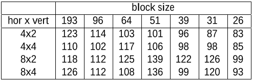
Table 1. Maximum Laplacian of a slice at ligamentum flavum for a human subject in set 1 (this analysis should be done for each set of ultrasound transducer parameters) 
Figure 1. Epidural needle insertion, midline approach. The epidural space is circled. 
Table 2. Gradient and Laplacian at regions of interest for human subjects for several methods 
Figure 3. Images for subject 15 and 5; the region in the white rectangle is used for quantitative evaluation of features near the epidural space, the structure being pointed to is the ligamentum flavum doublet and the circled structure is the lamina or bone boundary. 
Table 4. Computational cost of spatial compounding with warping for different parts of the algorithm. CPU time is calculated using a P4 3.0GHz with 1GB RAM, and a warping search region of ±4×±4 (set 2 with image size 636 x 359) and ±8×±2 (set 1 with image size 726 x 423) pixels. 
Figure 2. a) Ultrasound image of the lumbar spine with speckle noise and shadowing. A sagittal paramedian plane is used. b) Outlines of the reference and two beam steered images are shown. Spatial compounding uses positive and negative isonation angles to produce a set of beam-steered images that are subsequently combined. 
Figure 4. Images for subject 15 after compounding. The epidural space is the short horizontal line-pair near the middle. a) reference image, b) simple compounding, c) compounding with warping, d) compounding with warping and LP2+, e) median-based compounding with warping, f) median-based compounding with warping and LP2+. 
Figure 5. Images for subject 17 after compounding. The epidural space is the short horizontal line-pair at the bottom-left. a) reference image, b) simple compounding, c) compounding with warping, d) compounding with warping and LP2+, e) median-based compounding with warping, f) median-based compounding with warping and LP2+.


Citations
56 citations
Cites background from "Adaptive spatial compounding for im..."
...The possible correlation with fat thickness was initially thought to be helpful with automatic image processing of the features to emphasize the epidural space.(13,14) This is being investigated further because speed of sound variations can cause image distortion through compression and refraction....
[...]
13 citations
11 citations
2 citations
References
51 citations
45 citations
25 citations
"Adaptive spatial compounding for im..." refers background or methods in this paper
...A popular method is a coarse to fine, or multi-resolution approach where lower resolution blocks are registered and then, high resolution blocks are registered using a smaller search region[10]....
[...]
...Re-alignment of the features using an additional non-rigid registration (warping) was previously proposed to properly align the structures of each image[10]....
[...]
...We will choose images from −8o to 8 with a step size of 2 as it is a good tradeoff between individual image quality and number of images to average[10]....
[...]
...Averaging, also known as compounding, has been used previously in three ways: temporal compounding, spatial compounding [10][11][12] and frequency compounding[13]....
[...]
...The algorithm used for the course of this work builds on the work found in[10]....
[...]
7 citations
5 citations
"Adaptive spatial compounding for im..." refers methods in this paper
...The Canny edge detection technique[30] is a gradient-based method and is currently the most commonly used method as no methods have shown to consistently yield better performance[29][31][32][33]....
[...]