scispace - formally typeset
Search or ask a question
Journal ArticleDOI

Association of low back pain with muscle stiffness and muscle mass of the lumbar back muscles, and sagittal spinal alignment in young and middle-aged medical workers.

TL;DR: The results of this study suggest that LBP is associated with muscle stiffness of the lumbar multifidus muscle in young and middle‐aged medical workers.
About: This article is published in Clinical Biomechanics.The article was published on 2017-11-01 and is currently open access. It has received 61 citations till now. The article focuses on the topics: Muscle stiffness & Multifidus muscle.

Summary (3 min read)

1. Introduction

  • The occurrence rate of low back pain (LBP) within the lifetime of adults is about 80% (Waddell, 1987).
  • LBP occurs at a high occurrence rate in medical workers.
  • The relation of these electromyographic data with muscle stiffness is unknown.
  • The shear elastic modulus, as an index of muscle stiffness, is evaluated by measuring the shear wave propagation speed in the tissues that is generated by an ultrasonic SWE.
  • A decreased muscle mass in the lumbar erector spinae and quadratus lumborum muscles is also associated with LBP occurrence (Kamaz et al., 2007; Lee et al., 2011).

2.1. Participants

  • Thirty-two young and middle-aged medical workers in Kyoto Hakuaikai Hospital, Japan were included in the study.
  • The LBP group consisted of subjects with bilateral or central LBP (except for unilateral LBP) with a severity rating of ≥3 on the numerical rating scale (NRS) in both static (i.e., lying, sitting, or 6 standing) and dynamic situations (i.e., moving or walking), lasting 3 months or more at the time of evaluation.
  • Medical workers included nurses, care workers, and therapists.
  • The protocol was approved by the Ethics Committee of the Kyoto University Graduate School and Faculty of Medicine.

2.2. Low back pain assessment

  • The duration and degree of LBP, as well as the disabilities of daily living due to LBP, were assessed in the LBP group using a questionnaire.
  • The degree of LBP was examined using the NRS in both static (i.e., lying, sitting, or standing) and dynamic situations (i.e., moving or walking).
  • The disabilities of daily living due to LBP were assessed using the Oswestry disability index (ODI) (Fairbank and Pynsent, 2000).
  • The item of sex life, which can be removed if not applicable, was not used in this study.
  • The sum of each item was expressed as a percentage, and a large percentage indicated the severe disabilities of daily living due to LBP.

2.3. Ultrasound measurement

  • Images of the lumbar back muscles were taken using an ultrasound imaging device with SWE (Aixplorer, Supersonic Imagine, Aix-en-Provence, France).the authors.
  • Ultrasound images were measured once bilaterally for muscle thickness.
  • A previous study showed that the degree of intrarater reliability of the ultrasound technique is high for measuring muscle thickness of the lumbar back muscles (Masaki et al., 2015).
  • A linear array probe was set parallel to the muscle fibers to measure the shear elastic modulus accurately (Eby et al., 2013).
  • Three ROIs with a diameter of 10 mm were set in the color-coded box, with 1 located at the center of the box and the other 2 beside the initial ROI.

2.4. Measurement of spinal alignment

  • The Spinal Mouse (Index Ltd., Tokyo, Japan) was used to measure sagittal spinal alignment in the standing position (thoracic kyphosis, lumbar lordosis, and sacral anterior inclination angle) based on a previous study (Masaki et al., 2015).
  • Spinal alignment in the prone position was also measured to identify whether muscle stiffness and muscle mass of the lumbar back muscles were influenced by spinal alignment in the position of ultrasound measurement.
  • The Spinal Mouse was guided along the midline of the spine, starting at the C7 spinous process and ending at S3.
  • The lumbar lordosis angle was calculated from the sum of the 6 segmental angles from Th12/L1 to L5/S1.
  • The sacral anterior inclination angle was calculated from the difference between the sacral angle and the vertical plane.

2.5. Statistical analyses

  • Statistical analyses were performed using SPSS version 22.0 (IBM Japan; Tokyo, Japan).
  • Intraclass correlation coefficients [ICCs (1.1), ICC (1.2)] were calculated to examine intrarater reliabilities of the shear elastic modulus measurements.
  • Furthermore, the associations with LBP were investigated by multiple logistic regression analysis with a forward selection method.
  • This analysis was conducted using the shear elastic modulus and muscle thickness of the lumbar back muscles, and sagittal spinal alignment, age, body height, body weight, and sex as independent variables.
  • P values of <0.05 were considered significant.

3. Results

  • Table 1 presents characteristics and LBP status in the CTR and LBP groups.
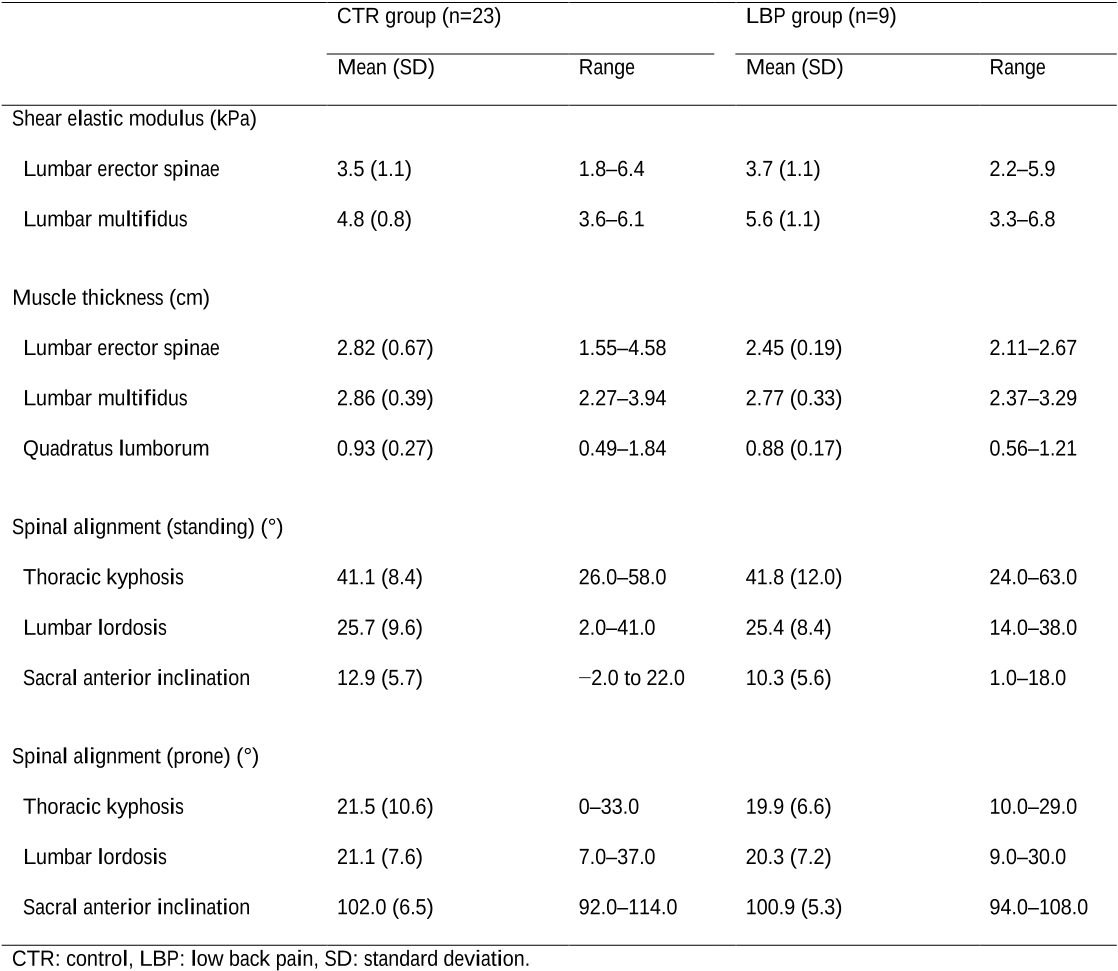
  • Muscle stiffness and muscle mass of the lumbar back muscle, and spinal alignment are shown in Table 2.
  • The shear elastic modulus of the lumbar multifidus muscle in the LBP group was significantly higher than that in the CTR group.
  • The height in the LBP group was significantly lower than that in the CTR group (Table 3).
  • Multiple logistic regression analysis also showed that the other factors were not significant independent determinants of LBP.

4. Discussion

  • Practicing rehabilitation based on the cause of LBP occurrence is important because the cause is attributed to different factors.
  • Muscle stiffness of the lumbar multifidus muscle is assumed to increase by muscle contraction.
  • Previous study (Chan et al., 2012) demonstrated muscle stiffness of the lumbar back muscles using a strain imaging method of the ultrasound imaging device, which is different from ultrasonic SWE used in the present study.the authors.
  • They targeted only the lumbar multifidus muscles in the LBP patients and suggested that no significant difference exists in the muscle stiffness of the lumbar multifidus muscle in the prone position between healthy subjects and LBP patients.
  • The results of the present study were consistent with those of the previous studies that demonstrate no association with spinal alignment in the standing position and LBP.

5. Conclusions

  • The results of the present study suggest that LBP is associated with muscle stiffness of the lumbar multifidus muscle rather than muscle stiffness of the lumbar erector spinae muscle, muscle mass of the lumbar back muscles, or sagittal spinal alignment in young and middle-aged medical workers.
  • No funding sources and potential conflicts of interest were disclosed for the present study.

Did you find this useful? Give us your feedback

Citations
More filters
Journal ArticleDOI
TL;DR: In patients with chronic radiculopathy, disc herniation and severe facet degeneration were associated with altered paraspinal muscle morphology at or below the pathology level, and recommendations are provided to promote uniform measurement techniques for future studies.
Abstract: Individual study results have demonstrated unclear relationships between neurocompressive disorders and paraspinal muscle morphology. This systematic review aimed to synthesize current evidence regarding the relationship lumbar neurocompressive disorders may have with lumbar paraspinal muscle morphology. Searches were conducted in seven databases from inception through October 2017. Observational studies with control or comparison groups comparing herniations, facet degeneration, or canal stenosis to changes in imaging or biopsy-identified lumbar paraspinal muscle morphology were included. Data extraction and risk of bias assessment were performed by review author pairs independent of one another. Morphological differences between individuals with and without neurocompressive disorders were compared qualitatively, and where possible, standardised mean differences were obtained. Twenty-eight studies were included. Lumbar multifidus fiber diameter was smaller on the side of and below herniation for type I [SMD: −0.40 (95% CI = −0.70, −0.09) and type II fibers [SMD: −0.38 (95% CI = −0.69, −0.06)] compared to the unaffected side. The distribution of type I fibers was greater on the herniation side [SMD: 0.43 (95% CI = 0.03, 0.82)]. Qualitatively, two studies assessing small angular fiber frequency and fiber type groupings demonstrated increases in these parameters below the herniation level. For diagnostic imaging meta-analyses, there were no consistent differences across the various assessment types for any paraspinal muscle groups when patients with herniation served as their own control. However, qualitative synthesis of between-group comparisons reported greater multifidus and erector spinae muscle atrophy or fat infiltration among patients with disc herniation and radiculopathy in four of six studies, and increased fatty infiltration in paraspinal muscles with higher grades of facet joint degeneration in four of five studies. Conflicting outcomes and variations in study methodology precluded a clear conclusion for canal stenosis. Based on mixed levels of risk of bias data, in patients with chronic radiculopathy, disc herniation and severe facet degeneration were associated with altered paraspinal muscle morphology at or below the pathology level. As the variability of study quality and heterogeneous approaches utilized to assess muscle morphology challenged comparison across studies, we provide recommendations to promote uniform measurement techniques for future studies. PROSPERO 2015: CRD42015012985

53 citations

Journal ArticleDOI
TL;DR: The myotonometer demonstrated acceptable reliability when used in a clinical setting in young adults with chronic LBP, and Measurements of the upper lumbar levels were not as reliable as those of the lower lumbAR levels.
Abstract: The reliability of a handheld myotonometer when used in a clinical setting to assess paraspinal muscle mechanical properties is unclear. This study aimed to investigate the between-session intra-rater reliability of a handheld myotonometer in young adults with low back pain (LBP) in a clinical environment. One assessor recorded lumbar paraspinal muscle tone and stiffness in an outpatient department on two occasions. The intraclass correlation coefficient (ICC), standard error of measurement (SEM), smallest real difference (SRD) and Bland-Altman analysis were conducted to assess reliability. The results indicated acceptable between-days intra-rater reliability (ICC > 0.75) for all measurements. The SEM of the muscle tone and stiffness measurements ranged between 0.20–0.66 Hz and 7.91–16.51 N/m, respectively. The SRD was 0.44–1.83 Hz for muscle tone and 21.93–52.87 N/m for muscle stiffness. SEM and SRD at L1-L2 were higher than those at other levels. The magnitude of agreement appeared to decrease as muscle tone and stiffness increased. The myotonometer demonstrated acceptable reliability when used in a clinical setting in young adults with chronic LBP. Measurements of the upper lumbar levels were not as reliable as those of the lower lumbar levels. The crural attachment of the diaphragm at L1 and L2 may affect paraspinal muscle tone and stiffness during respiratory cycles.

42 citations

Journal ArticleDOI
TL;DR: A differentiation in passive muscular stiffness between SM and DM is supported and evidence for an alteration in muscular stiffness at rest in individuals with LBP is provided to reflect a deficit in activation of the multifidus.

30 citations

Journal ArticleDOI
TL;DR: Resting lumbar muscle stiffness is greater in individuals with LBP than asymptomatic controls and is associated with self-reported pain and disability, but not physical exam findings.
Abstract: Background Lumbar muscle dysfunction is commonly implicated in low back pain (LBP). Shear-wave elastography (SWE) uses ultrasound technology to quantify absolute soft tissue stiffness (shear modulus), thereby allowing for estimation of individual muscle contraction and function. Objectives To compare resting and contracted stiffness of lumbar spine musculature in individuals with and without LBP using SWE. A secondary aim was to explore for relationships between common self-report and physical examination measures and resting and contracted muscle stiffness in individuals with LBP. Design Cross-sectional. Methods Shear modulus of the lumbar musculature was measured in 60 participants with LBP and 60 asymptomatic controls (120 total) using SWE. The lumbar erector spinae were imaged at rest only, while the lumbar multifidus was imaged at rest and during contraction. Before imaging, participants with LBP underwent a standardized clinical examination including a brief history, self-report questionnaires, and a physical examination. Lumbar muscle shear modulus was compared between participants with LBP and asymptomatic controls using ANCOVA. Potential associations between shear modulus and selected self-report and physical examination measures were assessed using correlation analysis. Results Stiffness of the erector spinae and lumbar multifidus at rest (but not during contraction) was greater in participants with LBP than in asymptomatic controls (p Conclusion Resting lumbar muscle stiffness is greater in individuals with LBP than asymptomatic controls and is associated with self-reported pain and disability, but not physical exam findings.

28 citations

Journal ArticleDOI
TL;DR: It is indicated that short-duration tissue manipulation can be an effective active break between prolonged sitting periods to prevent musculoskeletal issues, such as musculOSkeletal discomfort and back pain.

26 citations

References
More filters
Journal ArticleDOI
15 Nov 2000-Spine
TL;DR: The ODI remains a valid and vigorous measure and has been a worthwhile outcome measure, and the process of using the ODI is reviewed and should be the subject of further research.
Abstract: Study design The Oswestry Disability Index (ODI) has become one of the principal condition-specific outcome measures used in the management of spinal disorders. This review is based on publications using the ODI identified from the authors' personal databases, the Science Citation Index, and hand searches of Spine and current textbooks of spinal disorders. Objectives To review the versions of this instrument, document methods by which it has been validated, collate data from scores found in normal and back pain populations, provide curves for power calculations in studies using the ODI, and maintain the ODI as a gold standard outcome measure. Summary of background data It has now been 20 years since its original publication. More than 200 citations exist in the Science Citation Index. The authors have a large correspondence file relating to the ODI, that is cited in most of the large textbooks related to spinal disorders. Methods All the published versions of the questionnaire were identified. A systematic review of this literature was made. The various reports of validation were collated and related to a version. Results Four versions of the ODI are available in English and nine in other languages. Some published versions contain misprints, and many omit the scoring system. At least 114 studies contain usable data. These data provide both validation and standards for other users and indicate the power of the instrument for detecting change in sample populations. Conclusions The ODI remains a valid and vigorous measure and has been a worthwhile outcome measure. The process of using the ODI is reviewed and should be the subject of further research. The receiver operating characteristics should be explored in a population with higher self-report disabilities. The behavior of the instrument is incompletely understood, particularly in sensitivity to real change.

4,482 citations


"Association of low back pain with m..." refers methods in this paper

  • ...The disabilities of daily living due to LBP were assessed using the Oswestry disability index (ODI) (Fairbank and Pynsent, 2000)....

    [...]

  • ...The ODI consists of items, such as pain intensity of LBP during personal care (i.e., washing or dressing etc.), lifting, walking, sitting, standing, sleeping, sex life, social life, and travelling....

    [...]

Journal ArticleDOI
TL;DR: The present work focuses on the upright standing posture with different degree of lumbar lordosis, and by reduction of the number of unknown forces, a unique determination of the total force distributions at static equilibrium is obtained.
Abstract: From the mechanical point of view the spinal system is highly complex, containing a multitude of components, passive and active. In fact, even if the active components (the muscles) were exchanged by passive springs, the total number of elements considerably exceeds the minimum needed to maintain static equilibrium. In other words, the system is statically highly indeterminate. The particular role of the active components at static equilibrium is to enable a virtually arbitrary choice of posture, independent of the distribution and magnitude of the outer load albeit within physiological limits. Simultaneously this implies that ordinary procedures known from the analysis of mechanical systems with passive components cannot be applied. Hence the distribution of the forces over the different elements is not uniquely determined. Consequently nervous control of the force distribution over the muscles is needed, but little is known about how this achieved. This lack of knowledge implies great difficulties at numerical simulation of equilibrium states of the spinal system. These difficulties remain even if considerable reductions are made, such as the assumption that the thoracic cage behaves like a rigid body. A particularly useful point of view about the main principles of the force distributions appears to be the distinction between a local and a global system of muscles engaged in the equilibrium of the lumbar spine. The local system consists of muscles with insertion or origin (or both) at lumbar vertebrae, whereas the global system consists of muscles with origin on the pelvis and insertions on the thoracic cage. Given the posture of the lumbar spine, the force distribution over the local system appears to be essentially independent of the outer load of the body (though the force magnitudes are, of course, dependent on the magnitude of this load). Instead different distributions of the outer load on the body are met by different distributions of the forces in the global system. Thus, roughly speaking, the global system appears to take care of different distributions of outer forces on the body, whereas the local system performs an action, which is essentially locally determined (i.e. by the posture of the lumbar spine). The present work focuses on the upright standing posture with different degree of lumbar lordosis. The outer load is assumed to consist of weights carried on the shoulders. By reduction of the number of unknown forces, which is done by using a few different principles, a unique determination of the total force distributions at static equilibrium is obtained.(ABSTRACT TRUNCATED AT 400 WORDS)

1,241 citations


"Association of low back pain with m..." refers background in this paper

  • ...On the other hand, the lumbar multifidus muscle, which is a deep muscle of the trunk, is advantageous to stabilize the lumbar spine (Bergmark, 1989; MacDonald et al., 2006)....

    [...]

Journal ArticleDOI
Gordon Waddell1
01 Sep 1987-Spine
TL;DR: Observations of natural history and epidemiology suggest that low-back pain should be a benign, self-limiting condition, that low back-disability as opposed to pain is a relatively recent Western epidemic, and that the role of medicine in that epidemic must be critically examined.
Abstract: Because there is increasing concern about low-back disability and its current medical management, this analysis attempts to construct a new theoretic framework for treatment. Observations of natural history and epidemiology suggest that low-back pain should be a benign, self-limiting condition, that low back-disability as opposed to pain is a relatively recent Western epidemic, and that the role of medicine in that epidemic must be critically examined. The traditional medical model of disease is contrasted with a biopsychosocial model of illness to analyze success and failure in low-back disorders. Studies of the mathematical relationship between the elements of illness in chronic low-back pain suggest that the biopsychosocial concept can be used as an operational model that explains many clinical observations. This model is used to compare rest and active rehabilitation for low-back pain. Rest is the commonest treatment prescribed after analgesics but is based on a doubtful rationale, and there is little evidence of any lasting benefit. There is, however, little doubt about the harmful effects--especially of prolonged bed rest. Conversely, there is no evidence that activity is harmful and, contrary to common belief, it does not necessarily make the pain worse. Experimental studies clearly show that controlled exercises not only restore function, reduce distress and illness behavior, and promote return to work, but actually reduce pain. Clinical studies confirm the value of active rehabilitation in practice. To achieve the goal of treating patients rather than spines, we must approach low-back disability as an illness rather than low-back pain as a purely physical disease. We must distinguish pain as a purely the symptoms and signs of distress and illness behavior from those of physical disease, and nominal from substantive diagnoses. Management must change from a negative philosophy of rest for pain to more active restoration of function. Only a new model and understanding of illness by physicians and patients alike makes real change possible.

1,116 citations


"Association of low back pain with m..." refers background in this paper

  • ...The occurrence rate of low back pain (LBP) within the lifetime of adults is about 80% (Waddell, 1987)....

    [...]

Journal ArticleDOI
01 Dec 1996-Spine
TL;DR: Multifidus muscle recovery is not spontaneous on remission of painful symptoms, and lack of localized, muscle support may be one reason for the high recurrence rate of low back pain following the initial episode.
Abstract: Study Design. A clinical study was conducted on 39 patients with acute, first-episode, unilateral low back pain and unilateral, segmental inhibition of the multifidus muscle. Patients were allocated randomly to a control or treatment group. Objectives. To document the natural course of lumbar multifidus recovery and to evaluate the effectiveness of specific, localized, exercise therapy on muscle recovery. Summary of Background Data. Acute low back pain usually resolves spontaneously, but the recurrence rate is high. Inhibition of multifidus occurs with acute, first-episode, low back pain, and pathologic changes in this muscle have been linked with poor outcome and recurrence of symptoms. Methods. Patients in group 1 received medical treatment only. Patients in group 2 received medical treatment and specific, localized, exercise therapy. Outcome measures for both groups included 4 weekly assessments of pain, disability, range of motion, and size of the multifidus cross-sectional area. Independent examiners were blinded to group allocation. Patients were reassessed at a 10-week follow-up examination. Results. Multifidus muscle recovery was not spontaneous on remission of painful symptoms in patients in group 1. Muscle recovery was more rapid and more complete in patients in group 2 who received exercise therapy (P = 0.0001). Other outcome measurements were similar for the two groups at the 4-week examination. Although they resumed normal levels of activity, patients in group 1 still had decreased multifidus muscle size at the 10-week follow-up examination. Conclusions. Multifidus muscle recovery is not spontaneous on remission of painful symptoms. Lack of localized, muscle support may be one reason for the high recurrence rate of low back pain following the initial episode.

1,089 citations


"Association of low back pain with m..." refers background in this paper

  • ...Decreased muscle mass of the lumbar multifidus muscle (Barker et al., 2004; Cooper et al., 1992; Hides et al., 1996, 2008; Hodges et al., 2006; Keller et al., 2004) or changes in spinal alignment, such as decreased lumbar lordosis in the standing position (Tsuji et al., 2001), cause stress on…...

    [...]

Journal ArticleDOI
TL;DR: A comparative study between unoperated CLBP patients and matched control subjects with regard to muscle cross-sectional area (CSA) and the amount of fat deposits at different levels showed that only the CSA of the multifidus and only at the lowest level was found to be statistically smaller in LBP patients.
Abstract: Increasing documentation on the size and appearance of muscles in the lumbar spine of low back pain (LBP) patients is available in the literature. However, a comparative study between unoperated chronic low back pain (CLBP) patients and matched (age, gender, physical activity, height and weight) healthy controls with regard to muscle cross-sectional area (CSA) and the amount of fat deposits at different levels has never been undertaken. Moreover, since a recent focus in the physiotherapy management of patients with LBP has been the specific training of the stabilizing muscles, there is a need for quantifying and qualifying the multifidus. A comparative study between unoperated CLBP patients and matched control subjects was conducted. Twenty-three healthy volunteers and 32 patients were studied. The muscle and fat CSAs were derived from standard computed tomography (CT) images at three different levels, using computerized image analysis techniques. The muscles studied were: the total paraspinal muscle mass, the isolated multifidus and the psoas. The results showed that only the CSA of the multifidus and only at the lowest level (lower end-plate of L4) was found to be statistically smaller in LBP patients. As regards amount of fat, in none of the three studied muscles was a significant difference found between the two groups. An aetiological relationship between atrophy of the multifidus and the occurrence of LBP can not be ruled out as a possible explanation. Alternatively, atrophy may be the consequence of LBP: after the onset of pain and possible long-loop inhibition of the multifidus a combination of reflex inhibition and substitution patterns of the trunk muscles may work together and could cause a selective atrophy of the multifidus. Since this muscle is considered important for lumbar segmental stability, the phenomenon of atrophy may be a reason for the high recurrence rate of LBP.

594 citations


"Association of low back pain with m..." refers background in this paper

  • ...…imaging demonstrated that the muscle mass of the lumbar back muscles, such as the lumbar erector spinae, multifidus, and quadratus lumborum muscles, either decreases (Kamaz et al., 2007; Lee et al., 2011; Wallwork et al., 2009) or does not decrease (Danneels et al., 2000) in the LBP patients....

    [...]

  • ..., 2009) or does not decrease (Danneels et al., 2000) in the LBP patients....

    [...]

Frequently Asked Questions (11)
Q1. What contributions have the authors mentioned in the paper "Association of low back pain with muscle stiffness and muscle mass of the lumbar back muscles, and sagittal spinal alignment in young and middle-aged medical workers" ?

This study aimed to examine the association of LBP with muscle stiffness assessed using ultrasonic shear wave elastography ( SWE ) and muscle mass of the lumbar back muscle, and spinal alignment in young and middle-aged medical workers. The study comprised 23 asymptomatic medical workers [ control ( CTR ) group ] and 9 medical workers with LBP ( LBP group ). The association with LBP was investigated by multiple logistic regression analysis with a forward selection method. The results of this study suggest that LBP is associated with muscle stiffness of the lumbar multifidus muscle in young and middle-aged medical workers. 

In this case, the overuse caused by muscle spasm of the lumbar multifidus muscle may lead to circulatory difficulty within the muscle, which contributes to secondary LBP occurrence in the future. Further studies should examine training for improving muscle stiffness of the lumbar multifidus muscle effectively. They targeted only the lumbar multifidus muscles in the LBP patients and suggested that no significant difference exists in the muscle stiffness of the lumbar multifidus muscle in the prone position between healthy subjects and LBP patients. The present study suggests that LBP is associated with muscle stiffness of the lumbar multifidus muscle in young and middle-aged medical workers. 

which is caused from stress on intervertebral disks or intervertebral joints, may induce muscle spasm of the lumbar multifidus muscle. 

Circulatory difficulty within the lumbar multifidus muscle caused by overuse during the motions may contribute to an increase in muscle stiffness and LBP occurrence. 

Three ROIs with a diameter of 10 mm were set in the color-coded box, with 1 located at the center of the box and the other 2 beside the initial ROI. 

A possible reason for the association of LBP with muscle stiffness of the lumbar back muscles in the prone position is the frequent trunk flexion or pelvic anterior tilt of the medical workers in the standing position during medical treatment, care, and rehabilitation, as well as frequent extension of their trunk during transferring patients. 

The determination of the ROIs and the computation of muscle thickness and shear elastic modulus were performed by 1 examiner who was blinded to information of the groups. 

This inconsistency may be attributed to the flexion of the lumbar spine, which compensatorily becomes excessive during12medical treatment, care, rehabilitation, and transferring patients in medical workers, who have shorter body height (i.e., shorter upper extremities). 

shorter body height may contribute to increased muscle stiffness of the lumbar multifidus muscles, or the stress on intervertebral disks or intervertebral joints. 

The shear elastic modulus (G) was computed from the shear wave propagation speed (v) and the muscle mass density (ρ) using the following equation: G=ρv2 where ρ is presumed to be 1000 kg/m3 (Aubry et al., 2013). 

whether an increase in muscle stiffness of the lumbar multifidus muscle is caused by overuse or muscle spasm is unclear because the activities of the lumbar back muscles were not measured using electromyography during ultrasound measurement. 

Trending Questions (1)
What is association of low back pain muscle waekness?

The provided paper does not mention the association of low back pain with muscle weakness. The paper focuses on the association of low back pain with muscle stiffness and muscle mass of the lumbar back muscles, as well as sagittal spinal alignment in young and middle-aged medical workers.