scispace - formally typeset
Search or ask a question
Posted ContentDOI

Endothelial Pannexin 1-TRPV4 channel signaling lowers pulmonary arterial pressure

TL;DR: In this paper, the role of PANX1 in resistance-sized pulmonary arteries (PAs) is unknown. But, the authors hypothesized that PANx1-ATP-TRPV4 channel signaling promotes vasodilation and lowers pulmonary arterial pressure (PAP).
Abstract: Pannexin 1 (Panx1) is an ATP-efflux channel that controls endothelial function in the systemic circulation. However, the roles of endothelial Panx1 in resistance-sized pulmonary arteries (PAs) are unknown. Extracellular ATP dilates PAs through activation of endothelial TRPV4 (transient receptor potential vanilloid 4) ion channels. We hypothesized that endothelial Panx1–ATP–TRPV4 channel signaling promotes vasodilation and lowers pulmonary arterial pressure (PAP). Endothelial, but not smooth muscle, knockout of Panx1 or TRPV4 increased PA contractility and raised PAP. Panx1-effluxed extracellular ATP signaled through purinergic P2Y2 receptor (P2Y2R) to activate protein kinase Cα (PKCα), which in turn activated endothelial TRPV4 channels. Finally, caveolin-1 provided a signaling scaffold for endothelial Panx1, P2Y2R, PKCα, and TRPV4 channels in PAs, promoting their spatial proximity and enabling signaling interactions. These results indicate that endothelial Panx1–P2Y2R–TRPV4 channel signaling, facilitated by caveolin-1, reduces PA contractility and lowers PAP.

Summary (4 min read)

Introduction

  • The pulmonary endothelium exerts a dilatory influence on small, resistance-sized pulmonary arteries (PAs) and thereby lowers pulmonary arterial pressure (PAP).
  • Purinergic receptor signaling is an essential regulator of pulmonary vascular function [10] [11] [12] [13] .
  • Previous studies in small PAs showed that eATP activates TRPV4EC channels through P2 purinergic receptors, although the precise P2 receptor subtype was not identified 9 .
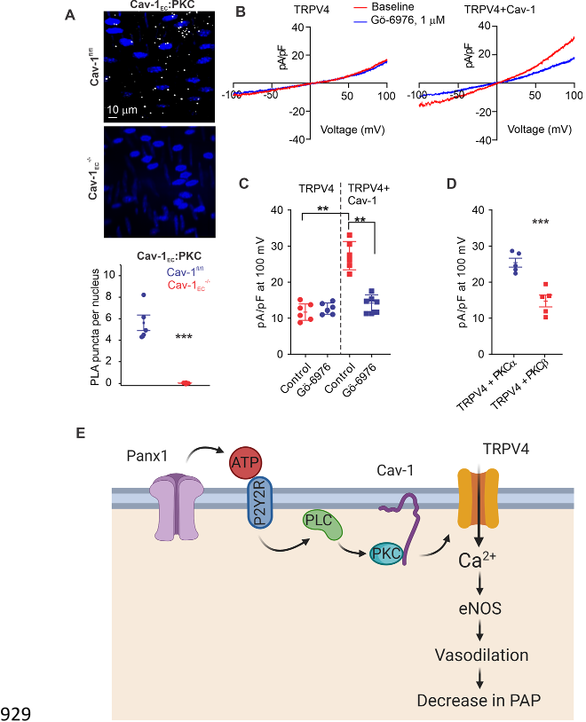
  • Here, the authors tested the hypothesis that Panx1EC-P2Y2REC-TRPV4EC channel signaling, supported by a signaling scaffold provided by Cav-1EC, reduces PA contractility and PAP.
  • -/mice, the authors show that endothelial Panx1-P2Y2R-TRPV4 signaling reduces PA contractility and lowers PAP.

Results

  • Endothelial, but not smooth muscle, Panx1-TRPV4 signaling lowers PA contractility.
  • In pressure myography experiments, ATP (1 µmol/L)-induced dilation was absent in PAs from TRPV4EC -/mice (Fig. 1B ), confirming that ATP dilates PAs through TRPV4EC channels.
  • Functional cardiac MRI studies indicated no alterations in cardiac function in TRPV4EC -/or Panx1EC -/mice compared with the respective control mice (Table 1 ), suggesting that the changes in RVSP were not due to altered cardiac function.
  • Localized, unitary Ca 2+ influx signals through TRPV4EC channels, termed TRPV4EC sparklets 25 , were recorded in en face, 4th-order PAs (~ 50 µm) loaded with Fluo-4.
  • These data support the concept that the reduced TRPV4EC channel activity in Panx1EC -/mice is due to impaired channel regulation rather than a decrease in the number of functional TRPV4EC channels.

Panx1EC-generated eATP acts through purinergic P2Y2REC stimulation to activate TRPV4EC channels.

  • Bioluminescence measurements confirmed lower baseline eATP levels in PAs from Panx1EC -/mice compared with PAs from Panx1 fl/fl mice (Fig. 2A ), supporting an essential role for Panx1EC channels as an eATP-release mechanism in PAs.
  • -/mice, P2Y2REC -/mice also showed elevated RVSP and an unaltered Fulton Index (Fig. 2G ).
  • Cav-1EC provides a scaffold for Panx1EC-P2Y2REC-TRPV4EC signaling.
  • Importantly, eATP-induced activation of TRPV4EC sparklets was absent in PAs from Cav-1EC -/mice (Fig. 3E ).

Cav-1EC anchoring of PKCα mediates P2Y2REC-dependent activation of TRPV4EC channels in PAs.

  • P2Y2R is a Gq protein-coupled receptor that activates the phospholipase C (PLC)diacylglycerol (DAG)-PKC signaling pathway.
  • Notably, PKC is known to phosphorylate TRPV4 channels and potentiate its activity 32 .
  • EATP, the DAG analog OAG (1 µmol/L), and the PKC activator phorbol myristate acetate (PMA; 10 nmol/L) stimulated TRPV4EC sparklet activity in small PAs (Fig. 4A-C ). Inhibition of PLC with U73122 (3 µmol/L) abolished eATP activation of TRPV4EC sparklets, but not OAG-or PMA-induced activation of TRPV4EC sparklets.
  • TRPV4EC channel activation by PLC-DAG-PKC signaling was further supported by increased activity of TRPV4EC sparklets in PAs from Cdh5-optoα1 adrenergic receptor (Cdh5-optoα1AR) mouse, which expresses lightsensitive α1AR in endothelial cells.
  • When activated with light (~473 nm), Optoα1AR generates the secondary messengers IP3 and diacylglycerol (DAG) 33 .

Discussion

  • Regulation of PA contractility and PAP is a complex process involving multiple cell types and signaling elements.
  • ECs from pulmonary capillaries and arteries are structurally and functionally different.
  • The authors data identify physiological roles of Panx1EC-TRPV4EC channel signaling in PAs, but whether such signaling operates in the capillary endothelium and is essential for its physiological function is unclear.
  • Furthermore, the activation of TRPV4EC channels by Panx1EC, eATP, P2Y2REC or PKCα requires Cav-1EC.
  • Further investigations are needed to determine whether impairment of this pathway contributes to elevated PAP in pulmonary vascular disorders and whether this pathway can be targeted for therapeutic benefit.

Drugs and chemical compounds.

  • Fluo-4-AM (Ca 2+ indicator) were purchased from Invitrogen (Carlsbad, CA, USA).
  • Tamoxifen and apyrase were obtained from Sigma-Aldrich ( St. Louis, MO, USA).

Animal protocols and models.

  • All animal protocols were approved by the University of Virginia Animal Care and Use Committee (protocols 4100 and 4120).
  • For surgical procedures, every effort was made to minimize suffering.
  • C57BL6/J were obtained from the Jackson Laboratory (Bar Harbor, ME).
  • Mice were used for experiments after a twoweek washout period.

Right ventricular systolic pressure (RVSP) and Fulton Index measurement.

  • Mice were anesthetized with pentobarbital (50 mg/kg bodyweight; intraperitoneally) and bupivacaine HCl (100 µL of 0.25% solution; subcutaneously) was used to numb the dissection site on the mouse.
  • RVSP was measured as an indirect indicator of pulmonary arterial pressure (PAP).
  • Right ventricular pressure and heart rate were acquired and analyzed using LabChart8 software (ADInstruments, Colorado Springs, CO).
  • A stable 3-minute recording was acquired for all the animals, and 1-minute continuous segment was used for data analysis.
  • When necessary, traces were digitally filtered using a low-pass filter at a cut-off frequency of 50 Hz.

Luciferase assay for total ATP release.

  • Isolated PAs were pinned down en face on a Sylgard block and cut open.
  • PAs were placed in black, opaque 96-well plates and incubated in HEPES-PSS for 10 minutes at 37 °C, followed by incubation with the ectonucleotidase inhibitor ARL 67156 (300 µmol/L, Tocris Bioscience, Minneapolis, MN) for 30 minutes at 37 °C.
  • 50 µL volume of each sample was transferred to another black, opaque 96-well plate.
  • ATP was measured using ATP bioluminescence assay reagent ATP Bioluminescence HSII kit (Roche Applied Science, Penzberg, Germany).
  • Using a luminometer (FluoStar Omega), 50 µL of luciferin:luciferase reagent (ATP bioluminescence assay kit HSII; Roche Applied Science, Penzberg, Germany) was injected into each well and luminescence was recorded following a 5 second orbital mix and sample measurement at 7 seconds.

Cardiac Magnetic Resonance Imaging (MRI).

  • MRI studies were conducted under protocols that comply with the Guide for the Care and Use of Laboratory Animals (NIH publication no.
  • Mice were positioned in the scanner under 1.25% isoflurane anesthesia and body temperature was maintained at 37°C using thermostatic circulating water.
  • MRI was performed on a 7 Tesla (T) Clinscan system (Bruker, Ettlingen, Germany) equipped with actively shielded gradients with a full strength of 650 mT/m and a slew rate of 6666 mT/m/ms 60 .
  • Six short-axis slices were acquired from base to apex, with slice thickness of 1 mm, in-plane spatial resolution of 0.2 × 0.2 mm 2 , and temporal resolution of 8-12 ms.
  • Baseline ejection fraction (EF), end-diastolic volume (EDV), end-systolic volume (ESV), myocardial mass, wall thickness, stroke volume (SV), and cardiac output (CO) were assessed from the cine images using the freely available software Segment version 2.0 R5292 (http://segment.heiberg.se).

Pressure myography.

  • All other pharmacological treatments were performed in the presence of U46619.
  • Before measurement of vascular reactivity, arteries were treated with NS309 (1 µmol/L), a direct opener of endothelial IK/SK channels, to assess endothelial health.
  • Percent constriction was calculated by: [(Diameterbefore−Diameterafter)/Diameterbefore] × 100 (1) where Diameterbefore is the diameter of the artery before a treatment and Diameterafter is the diameter after the treatment.

Calculation of TRPV4 sparklet activity per site.

  • Area under the curve for all the events at a site was determined using trapezoidal numerical integration ([F−F0]/F0 over time, in seconds).
  • NPO was determined using Single Channel Search module of Clampfit and quantal amplitudes derived from all-points histograms 9 (ΔF/F0 of 0.29 for Fluo-4 -loaded PAs).
  • Total number of sparklet sites in a field was divided by the number of cells in that field to obtain sparklet sites per cell.

Immunostaining.

  • PAs were fixed with 4% paraformaldehyde (PFA) at room temperature for 15 minutes and then washed 3 times with phosphate-buffered saline (PBS).
  • Following the overnight incubation, PAs were incubated with secondary antibody 1:500 Alexa Fluor® 568-conjugated donkey anti-rabbit (Life Technologies, Carlsbad, CA, USA) for one hour at room temperature in the dark room.
  • For nuclear staining, PAs were washed with PBS and then incubated with 0.3 mmol/L DAPI (Invitrogen, Carlsbad, CA, USA) for 10 minutes at room temperature.
  • Images were acquired along the z-axis from the surface of the endothelium to the bottom where the EC layer encounters the smooth muscle cell layer with a slice size of 0.1 µm using the Andor microscope described above.
  • Immunostaining for the protein of interest was evaluated using an excitation of 561 nm and collecting the emitted fluorescence with a 607/36 nm band-pass filter.

In situ Proximity Ligation Assay (PLA).

  • PAs were pinned en face on SYLGARD blocks.
  • PAs were then permeabilized with 0.2% Triton X for 30 minutes at room temperature followed by blocking with 5% normal donkey serum (Abcam plc, Cambridge, MA, USA) and 300 mmol/L glycine for one hour at room temperature.
  • The PLA protocol from Duolink PLA Technology kit (Sigma-Aldrich, St. Louis, MO, USA) was followed for the detection of co-localized proteins.
  • Lastly, PAs were incubated with 0.3 mmol/L DAPI nuclear staining (Invitrogen, Carlsbad, CA, USA) for 10 minutes at room temperature in the dark room.
  • PLA images were acquired using the Andor Revolution spinning-disk confocal imaging system along the z-axis at a slice size of 0.1 µm.

Plasmid generation and transfection into HEK293 cells.

  • The TRPV4 coding sequence without stop codons was amplified from mouse heart cDNA.
  • The amplified fragment was inserted into a plasmid backbone containing a CMV promoter region for expression and in addition, is suitable for lentiviral production by Gibson assembly.
  • The inframe FLAG tag was inserted into the 3'-primer used for amplification.
  • HEK293 cells were seeded (7 x 10 5 cells per 100 mm dish) in Dulbecco's Modified Eagle Medium with 10% fetal bovine serum (Thermo Fisher Scientific Inc., Waltham, MA, USA) 1 day prior to transfection.
  • TRPV4 was co-expressed with PKCα and PKCβ, obtained from Origene Technologies (Montgomery County, MD).

Patch clamp in HEK293 cells and freshly isolated ECs.

  • TRPV4 channel current was recorded in HEK293 cells using whole-cell patch configuration 48 hrs after transfection.
  • Outward currents were obtained by averaging the currents through the voltage step.
  • GSK219-sensitive currents were obtained by subtracting the currents in the presence of GSK219 from the currents in the presence of GSK101.

Did you find this useful? Give us your feedback

Figures (7)

Content maybe subject to copyright    Report

1
Endothelial Pannexin 1–TRPV4 channel signaling 1
lowers pulmonary arterial pressure 2
Panx1-TRPV4 signaling in pulmonary endothelium 3
4
1
Zdravka Daneva;
1,2
Matteo Ottolini;
1
Yen-Lin Chen;
1
Eliska Klimentova;
3
Soham A. Shah; 5
4
Richard D. Minshall;
5
Cheikh I. Seye,
6
Victor E. Laubach;
7
Brant E. Isakson;
1,7
Swapnil K. 6
Sonkusare 7
8
1
Robert M. Berne Cardiovascular Research Center, University of Virginia, Charlottesville, VA, 22908, 9
USA;
10
2
Department of Pharmacology, University of Virginia, Charlottesville, VA, 22908, USA; 11
3
Department of Biomedical Engineering, University of Virginia, Charlottesville, VA, 22908, USA; 12
4
Department of Anesthesiology, University of Illinois at Chicago, Chicago, IL, USA; Department of 13
Pharmacology, University of Illinois at Chicago, Chicago, IL, USA; 14
5
Department of Biochemistry, University of Missouri-Columbia, Columbia, MO, USA; 15
6
Department of Surgery, University of Virginia, Charlottesville, VA, 22908, USA; 16
7
Department of Molecular Physiology and Biological Physics, University of Virginia, Charlottesville, 17
VA, 22908, USA 18
19
20
Correspondence should be addressed to: 21
Swapnil K. Sonkusare, Ph.D. 22
University of Virginia School of Medicine 23
P.O. Box 801394 24
Charlottesville, VA 22908 25
E-mail: sks2n@virginia.edu 26
Phone: 434-297-7401 27
28
29
30
Key Words: Pannexin 1, TRP channels, pulmonary artery, endothelium, purinergic signaling, 31
caveolin 1. 32
33
.CC-BY 4.0 International licenseavailable under a
(which was not certified by peer review) is the author/funder, who has granted bioRxiv a license to display the preprint in perpetuity. It is made
The copyright holder for this preprintthis version posted March 9, 2021. ; https://doi.org/10.1101/2021.03.09.434532doi: bioRxiv preprint

2
Abstract. 34
Pannexin 1 (Panx1) is an ATP-efflux channel that controls endothelial function in the systemic 35
circulation. However, the roles of endothelial Panx1 in resistance-sized pulmonary arteries (PAs) 36
are unknown. Extracellular ATP dilates PAs through activation of endothelial TRPV4 (transient 37
receptor potential vanilloid 4) ion channels. We hypothesized that endothelial Panx1ATP38
TRPV4 channel signaling promotes vasodilation and lowers pulmonary arterial pressure (PAP). 39
Endothelial, but not smooth muscle, knockout of Panx1 or TRPV4 increased PA contractility and 40
raised PAP. Panx1-effluxed extracellular ATP signaled through purinergic P2Y2 receptor 41
(P2Y2R) to activate protein kinase Cα (PKCα), which in turn activated endothelial TRPV4 42
channels. Finally, caveolin-1 provided a signaling scaffold for endothelial Panx1, P2Y2R, PKCα, 43
and TRPV4 channels in PAs, promoting their spatial proximity and enabling signaling interactions. 44
These results indicate that endothelial Panx1P2Y2R–TRPV4 channel signaling, facilitated by 45
caveolin-1, reduces PA contractility and lowers PAP. 46
47
48
49
50
51
52
53
54
.CC-BY 4.0 International licenseavailable under a
(which was not certified by peer review) is the author/funder, who has granted bioRxiv a license to display the preprint in perpetuity. It is made
The copyright holder for this preprintthis version posted March 9, 2021. ; https://doi.org/10.1101/2021.03.09.434532doi: bioRxiv preprint

3
Introduction 55
The pulmonary endothelium exerts a dilatory influence on small, resistance-sized 56
pulmonary arteries (PAs) and thereby lowers pulmonary arterial pressure (PAP). However, 57
endothelial signaling mechanisms that control PA contractility remain poorly understood. In this 58
regard, pannexin 1 (Panx1), which is expressed in the pulmonary endothelium and epithelium
1
, 59
has emerged as a crucial controller of endothelial function
2, 3
. Panx1, the most studied member of 60
the pannexin family, forms a hexameric transmembrane channel at the cell membrane that allows 61
efflux of ATP from the cytosol
4, 5
. Previous studies have indicated that Panx1
EC
promotes 62
endothelium-dependent dilation of systemic arteries
6, 7
, and endothelial cell (EC) Panx1 (Panx1
EC
) 63
has been linked to inflammation in pulmonary capillaries
8
. Beyond this, however, the 64
physiological roles of Panx1
EC
in the pulmonary vasculature are largely unknown. 65
Extracellular ATP (eATP) was recently shown to activate TRPV4 (transient receptor 66
potential vanilloid 4) channels in the endothelium of small PAs
9
, establishing endothelial TRPV4 67
(TRPV4
EC
) channels as potential signaling targets of Panx1
EC
in the pulmonary circulation. Ca
2+
68
influx through TRPV4
EC
channels is known to dilate small PAs through activation of endothelial 69
nitric oxide synthase (eNOS)
9
. These observations suggest that Panx1
EC
-released eATP may act 70
through TRPV4
EC
channels to reduce PA contractility and lower PAP. 71
Purinergic receptor signaling is an essential regulator of pulmonary vascular function
10-13
. 72
Previous studies in small PAs showed that eATP activates TRPV4
EC
channels through P2 73
purinergic receptors, although the precise P2 receptor subtype was not identified
9
. Pulmonary 74
endothelium expresses both P2Y and P2X receptor subtypes. Konduri et al. showed that eATP 75
dilates PAs through P2Y2 receptor (P2Y2R) activation and subsequent endothelial NO release
13
. 76
Recent evidence from systemic ECs and other cell types also supports P2Y2R-dependent 77
.CC-BY 4.0 International licenseavailable under a
(which was not certified by peer review) is the author/funder, who has granted bioRxiv a license to display the preprint in perpetuity. It is made
The copyright holder for this preprintthis version posted March 9, 2021. ; https://doi.org/10.1101/2021.03.09.434532doi: bioRxiv preprint

4
activation of TRPV4 channels by eATP
14, 15
. These findings raise the possibility that the 78
endothelial P2Y2 receptor (P2Y2R
EC
) may be the signaling intermediate for Panx1
EC
TRPV4
EC
79
channel communication in PAs. 80
The linkage between Panx1
EC
-mediated eATP release and subsequent activation of 81
P2Y2R
EC
TRPV4
EC
signaling could depend on the spatial proximity of individual elements82
Panx1
EC
, P2Y2R
EC
, and TRPV4
EC
a functionality possibly provided by a signaling scaffold. 83
Caveolin-1 (Cav-1), a structural protein that interacts with and stabilizes other proteins in the 84
pulmonary circulation
16
, co-localizes with Panx1, P2Y2R, and TRPV4 channels in multiple cell 85
types
17-19
. Notably, global Cav-1
-/-
mice show elevated PAP, and endothelial Cav-1 (Cav-1
EC
)-86
dependent signaling is impaired in pulmonary hypertension
20-22
. 87
Here, we tested the hypothesis that Panx1
EC
–P2Y2R
EC
TRPV4
EC
channel signaling, 88
supported by a signaling scaffold provided by Cav-1
EC
, reduces PA contractility and PAP. Using 89
inducible, EC-specific Panx1
-/-
, TRPV4
-/-
, P2Y2R
-/-
and Cav-1
EC
-/-
mice, we show that endothelial 90
Panx1–P2Y2R–TRPV4
signaling reduces PA contractility and lowers PAP. Panx1
EC
-generated 91
eATP acts via P2Y2R
EC
stimulation to activate protein kinase Cα (PKCα) and thereby increase 92
TRPV4
EC
channel activity. Panx1
EC
, P2Y2R
EC
, PKCα, and TRPV4
EC
channels co-localize with 93
Cav-1
EC,
ensuring spatial proximity among the individual elements and supporting signaling 94
interactions. Overall, these findings advance our understanding of endothelial mechanisms that 95
control PAP and suggest the possibility of targeting these mechanisms to lower PAP in pulmonary 96
vascular disorders. 97
98
99
.CC-BY 4.0 International licenseavailable under a
(which was not certified by peer review) is the author/funder, who has granted bioRxiv a license to display the preprint in perpetuity. It is made
The copyright holder for this preprintthis version posted March 9, 2021. ; https://doi.org/10.1101/2021.03.09.434532doi: bioRxiv preprint

5
Results 100
Endothelial, but not smooth muscle, Panx1TRPV4
signaling lowers PA contractility. 101
To clearly define the physiological roles of Panx1
EC
and TRPV4
EC
channels, we utilized 102
tamoxifen-inducible, EC-specific Panx1
EC
-/-
and TRPV4
EC
-/-
mice
23, 24
. Tamoxifen-injected 103
TRPV4
fl/fl
Cre
-
(TRPV4
fl/fl
) or Panx1
fl/fl
Cre
-
(Panx1
fl/fl
) mice were used as controls
8, 23
. 104
TRPV4
EC
-/-
mice showed elevated right ventricular systolic pressure (RVSP), a commonly used 105
in vivo indicator of PAP (Fig. 1A). In pressure myography experiments, ATP (1 µmol/L)-induced 106
dilation was absent in PAs from TRPV4
EC
-/-
mice (Fig. 1B), confirming that ATP dilates PAs 107
through TRPV4
EC
channels. RVSP was also elevated in Panx1
EC
-/-
mice (Fig. 1C). The Fulton 108
Index, a ratio of right ventricular (RV) weight to left ventricle plus septal (LV + S) weight, was 109
not altered in TRPV4
EC
-/-
or Panx1
EC
-/-
mice compared with the respective control mice, suggesting 110
a lack of right ventricular hypertrophy in these mice (Table 1). Importantly, baseline RVSP was 111
not altered in inducible, SMC-specific TRPV4 (TRPV4
SMC
-/-
) or Panx1 (Panx1
SMC
-/-
) knockout 112
mice (Fig. 1A and C). Functional cardiac MRI studies indicated no alterations in cardiac function 113
in TRPV4
EC
-/-
or Panx1
EC
-/-
mice compared with the respective control mice (Table 1), suggesting 114
that the changes in RVSP were not due to altered cardiac function. 115
Localized, unitary Ca
2+
influx signals through TRPV4
EC
channels, termed TRPV4
EC
116
sparklets
25
, were recorded in en face
,
4th-order PAs (~ 50 µm) loaded with Fluo-4. Baseline 117
TRPV4
EC
sparklet activity and activity induced by a low concentration (1 nmol/L) of the specific 118
TRPV4 channel agonist, GSK1016790A (hereafter, GSK101), were significantly reduced in PAs 119
from Panx1
EC
-/-
mice compared with those from Panx1
fl/fl
mice (Fig. 1D). Additionally, the number 120
of TRPV4
EC
sparklet sites per cell was decreased in PAs from Panx1
EC
-/-
mice (Fig. 1E). At a 121
.CC-BY 4.0 International licenseavailable under a
(which was not certified by peer review) is the author/funder, who has granted bioRxiv a license to display the preprint in perpetuity. It is made
The copyright holder for this preprintthis version posted March 9, 2021. ; https://doi.org/10.1101/2021.03.09.434532doi: bioRxiv preprint

Citations
More filters
Journal ArticleDOI
TL;DR: This work shows that the endothelium resolves the issue of distributed sensing by using a network with scale-free and small-world properties, which confers a high signal-propagation speed and a high degree of synchronizability across the endot Helium.
Abstract: Significance The endothelium is the single layer of cells lining all blood vessels and acts as a central control hub to regulate multiple cardiovascular functions in response to hundreds of physiological stimuli. The detection of various physiological stimuli is distributed in spatially separated sites across the endothelium. Distributed sensing is difficult to reconcile with the requirement for coordinated cell activity across large regions of the endothelium. Here, we show that the endothelium resolves the issue by using a network with scale-free and small-world properties. The organization confers a high signal-propagation speed and a high degree of synchronizability across the endothelium. The network organization also explains the robust nature of endothelial communication and its resistance to damage or failure.

6 citations

Journal ArticleDOI
TL;DR: Depolarised mitochondria switch TRPV4 signalling from relying on Ca-induced Ca release at IP receptors, to being independent of Ca influx and instead mediated by ATP release via pannexins, highlighting a previously unknown role of mitochondria in shaping TRpV4 mediated Ca signalling by facilitating ATP release.
Abstract: Background and Purpose: Ca2+ influx via TRPV4 channels triggers Ca2+ release from the IP3‐sensitive internal store to generate repetitive oscillations. Although mitochondria are acknowledged regulators of IP3‐mediated Ca2+ release, how TRPV4‐mediated Ca2+ signals are regulated by mitochondria is unknown. We show that depolarised mitochondria switch TRPV4 signalling from relying on Ca2+‐induced Ca2+ release at IP3 receptors to being independent of Ca2+ influx and instead mediated by ATP release via pannexins. Experimental Approach: TRPV4‐evoked Ca2+ signals were individually examined in hundreds of cells in the endothelium of rat mesenteric resistance arteries using the indicator Cal520. Key Results: TRPV4 activation with GSK1016790A (GSK) generated repetitive Ca2+ oscillations that required Ca2+ influx. However, when the mitochondrial membrane potential was depolarised, by the uncoupler CCCP or complex I inhibitor rotenone, TRPV4 activation generated large propagating, multicellular, Ca2+ waves in the absence of external Ca2+. The ATP synthase inhibitor oligomycin did not potentiate TRPV4‐mediated Ca2+ signals. GSK‐evoked Ca2+ waves, when mitochondria were depolarised, were blocked by the TRPV4 channel blocker HC067047, the SERCA inhibitor cyclopiazonic acid, the PLC blocker U73122 and the inositol trisphosphate receptor blocker caffeine. The Ca2+ waves were also inhibited by the extracellular ATP blockers suramin and apyrase and the pannexin blocker probenecid. Conclusion and Implications: These results highlight a previously unknown role of mitochondria in shaping TRPV4‐mediated Ca2+ signalling by facilitating ATP release. When mitochondria are depolarised, TRPV4‐mediated release of ATP via pannexin channels activates plasma membrane purinergic receptors to trigger IP3‐evoked Ca2+ release.

5 citations

References
More filters
Journal ArticleDOI
TL;DR: Pannexin 1 channels have a wide expression spectrum, they are of large conductance and permeant for ATP, and they are mechanosensitive, Hence, pannexins are candidates for the release of ATP to the extracellular space upon mechanical stress.

739 citations

Journal ArticleDOI
23 Apr 2009-Nature
TL;DR: The development of a versatile family of genetically encoded optical tools that leverage common structure–function relationships among G-protein-coupled receptors (GPCRs) to recruit and control, with high spatiotemporal precision, receptor-initiated biochemical signalling pathways.
Abstract: In the study of complex mammalian behaviours, technological limitations have prevented spatiotemporally precise control over intracellular signalling processes. Here we report the development of a versatile family of genetically encoded optical tools ('optoXRs') that leverage common structure-function relationships among G-protein-coupled receptors (GPCRs) to recruit and control, with high spatiotemporal precision, receptor-initiated biochemical signalling pathways. In particular, we have developed and characterized two optoXRs that selectively recruit distinct, targeted signalling pathways in response to light. The two optoXRs exerted opposing effects on spike firing in nucleus accumbens in vivo, and precisely timed optoXR photostimulation in nucleus accumbens by itself sufficed to drive conditioned place preference in freely moving mice. The optoXR approach allows testing of hypotheses regarding the causal impact of biochemical signalling in behaving mammals, in a targetable and temporally precise manner.

706 citations

Journal ArticleDOI
TL;DR: It is shown that Gq-G11-mediated signaling in smooth muscle cells is required for maintenance of basal blood pressure and for the development of salt-induced hypertension, which identifies the G12-G13–LARG–mediated signaling pathway as a new target for antihypertensive therapies that would be expected to leave normal blood pressure regulation unaffected.
Abstract: The tone of vascular smooth muscle cells is a primary determinant of the total peripheral vascular resistance and hence the arterial blood pressure. Most forms of hypertension ultimately result from an increased vascular tone that leads to an elevated total peripheral resistance1,2,3. Regulation of vascular resistance under normotensive and hypertensive conditions involves multiple mediators, many of which act through G protein–coupled receptors on vascular smooth muscle cells4. Receptors that mediate vasoconstriction couple with the G-proteins Gq-G11 and G12-G13 to stimulate phosphorylation of myosin light chain (MLC) via the Ca2+/MLC kinase– and Rho/Rho kinase–mediated signaling pathways, respectively4,5,6. Using genetically altered mouse models that allow for the acute abrogation of both signaling pathways by inducible Cre/loxP-mediated mutagenesis in smooth muscle cells, we show that Gq-G11–mediated signaling in smooth muscle cells is required for maintenance of basal blood pressure and for the development of salt-induced hypertension. In contrast, lack of G12-G13, as well as of their major effector, the leukemia-associated Rho guanine nucleotide exchange factor (LARG), did not alter normal blood pressure regulation but did block the development of salt-induced hypertension. This identifies the G12-G13–LARG–mediated signaling pathway as a new target for antihypertensive therapies that would be expected to leave normal blood pressure regulation unaffected.

583 citations

Journal ArticleDOI
TL;DR: It is shown that pannexin 1 channels can be activated by extracellular ATP acting through purinergic receptors of the P2Y group as well as by cytoplasmic calcium.

500 citations

Journal ArticleDOI
04 May 2012-Science
TL;DR: Results support the concept that Ca2+ influx through single TRPV4 channels is leveraged by the amplifier effect of cooperative channel gating and the high Ca2- sensitivity of IK and SK channels to cause vasodilation.
Abstract: Major features of the transcellular signaling mechanism responsible for endothelium-dependent regulation of vascular smooth muscle tone are unresolved. We identified local calcium (Ca(2+)) signals ("sparklets") in the vascular endothelium of resistance arteries that represent Ca(2+) influx through single TRPV4 cation channels. Gating of individual TRPV4 channels within a four-channel cluster was cooperative, with activation of as few as three channels per cell causing maximal dilation through activation of endothelial cell intermediate (IK)- and small (SK)-conductance, Ca(2+)-sensitive potassium (K(+)) channels. Endothelial-dependent muscarinic receptor signaling also acted largely through TRPV4 sparklet-mediated stimulation of IK and SK channels to promote vasodilation. These results support the concept that Ca(2+) influx through single TRPV4 channels is leveraged by the amplifier effect of cooperative channel gating and the high Ca(2+) sensitivity of IK and SK channels to cause vasodilation.

454 citations

Related Papers (5)