scispace - formally typeset
Search or ask a question
Journal ArticleDOI

In vivo screening of mangrove plants for anti WSSV activity in Penaeus monodon, and evaluation of Ceriops tagal as a potential source of antiviral molecules

03 Feb 2011-Aquaculture (Elsevier)-Vol. 311, Iss: 1, pp 36-41
TL;DR: The study indicated suitability of the aqueous extract of C. tagal as a possible prophylaxis for WSSV infection in shrimp, the first report on the anti W SSV property of the mangrove plant C.tagal.
About: This article is published in Aquaculture.The article was published on 2011-02-03 and is currently open access. It has received 52 citations till now. The article focuses on the topics: Ceriops tagal & Penaeus monodon.

Summary (5 min read)

1. Introduction

  • White spot syndrome virus (WSSV), an enveloped non occluded DNA (300 kb) virus of the family Nimaviride under the new genus Wispovirus (Mayo, 2002), is the most devastating shrimp pathogen ever isolated and studied; it causes total mortality to a rearing stock within 3–7 days of infection in a culture system (Lightner, 1996).
  • The protective effect of Cynodon dactylyon (a terrestrial plant) in Penaeus monodon fromWSSV has also been documented (Balasubramanian et al., 2007;2008).
  • There has never been any report on the anti WSSV activity in mangrove plants.

2. Methods

  • 1. Collection and identification of mangrove plants Mangrove plants such as Excoecaria agallocha, Acanthus ilicifolius, Avicennia sp., Rhizophora mucronata, Rhizophora apiculata, Sonneratia sp. and Ceriops tagal were collected from different localities in South India (9° 58′ 1.20″N, 76° 15′ 0.00″E).
  • The plants were identified following Naskar and Mandal (1999), coded and voucher specimens deposited in the herbarium collection of the National Centre for Aquatic Animal Health, Cochin University of Science and Technology.

2.2. Aqueous extract from mangrove plants

  • Leaves were shade dried, powdered and used for the preparation of the aqueous extract using a protocol developed here.
  • Accordingly, 50 g of mangrove plant leaf powder was soaked in a minimum quantity of double distilled water and frozen to −20 °C, thawed and frozen repeatedly for three times and extracted to 500 ml final volume of double distilled water in a Warring blender at an ambient temperature.
  • These preparations were examined for their virucidal activity and for their protective effects fromWSSVon oral administration in P. monodon.

2.3. Virus inoculum

  • A composite sample of gills and soft parts of cephalothorax (500 mg) from freshly infected P.monodonwasmacerated in 10 ml cold PBS (NaCl 8 g, KCl 0.2 g, Na2HPO4 1.15 g, KH2PO4 0.2 g, double distilled water 100 ml)withglasswool to ahomogenous slurry usingmortar andpestle in an ice bath.
  • Viral infectivity titre was determined as the extractable virus and expressed as LD 50 in shrimp following Reed and Muench (1938).
  • The virus stock thus prepared for the experiment was stored in 500 μl aliquots at −80 °C.

2.4. Preparation of mangrove extract coated feed

  • The aqueous extracts were lyophilized and the dry mass re suspended in the required quantity of distilled water and coated onto feed pellets to arrive at a concentration of 1% w/w.
  • The above preparation was dried under vacuum and used for oral administration.

2.5. Experimental animals

  • All animals used in this studywere single spawner bred,WSSV free juveniles of P. monodon grown in a recirculating aquaculture system at the National Centre for Aquatic Animal Health.
  • The WSSV free status was confirmed through frequent Nested PCR analysis using a commercial kit (Bangalore Genei, Bangalore, India) of the shrimps during the culture operation in the recirculation system and also before the start of the experiment.
  • The shrimps weighing 4–5 g were maintained in 30 liter capacity fiber reinforced plastic (FRP) tanks with diluted sea water at salinity 15.
  • Uniformly 10% water was exchanged every day.

2.6. Virucidal activity of the aqueous extract

  • The aqueous plant extracts (0.5 ml) were mixed with equal volumes of viral suspension and incubated for 3 h at 25 °C.
  • The controls included mixtures of WSSV and PBS (positive control) and PBS alone (negative control).
  • From each of the preparation, aliquots of 10 μl each were intramuscularly administered to the animals (5×4 = 20 animals each) and monitored for 15 days.
  • Gill tissue was extracted from moribund animals and the controls, which survived the 15 day period of experimentation during the course of the experiment.
  • The samples were preserved in 70% ethanol for diagnostic PCR to detect WSSV.

2.7. Oral administration of the plant extracts along with diet and challenge with WSSV

  • All test animals (5×4=20 animals each) were fed with the plant extract coated feed at a rate of 10% of the bodyweight two times a day.
  • Feeding continued for 15 days, and the animals were challenged by feeding with freshly generatedWSSV infected tissue at a rate of 10% of the body weight, and kept under observation for 15 days on the respective diet (normal diet for the positive and negative controls and the diet coatedwith the plant extract in the test group).
  • Gill tissuewas extracted from moribund/dead animals and from those which survived the challenge with WSSV, and was preserved in 70% ethanol for diagnostic PCR.
  • On completion of the experiment, tissue homogenates were prepared from the test and control animals and passaged to a fresh batch of nested PCR negative animals as bio assay to check the presence of virions in the survived animals.
  • The presence of WSSV DNA was further examined by way of nested PCR.

2.9. Diagnostic PCR of the extracted tissue samples

  • For diagnostic PCR, DNA from the gill tissue was extracted in DNAzol according to the manufacturer's protocol.
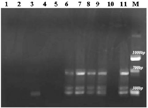
  • A WSSV nested PCR detection kit (Bangalore Geni) that yielded 650 and 300 bp WSSV specific amplicons was used for amplification of the viral DNA.
  • Following the instructions given with the kit, the amplified product was generated in a thermal cycler .
  • The PCR products were then analyzed on 1% W/V agarose gels using TAE (1X) buffer (Tris–HCL 0.04 M, EDTA 0.0001 M, Glacial acetic acid 5.71%), stained with ethidium bromide and visualized on a gel documentation system, Dolphin-Doc (Weal Tec, USA).

2.10. HPLC analysis of the crude aqueous extract from C. tagal

  • On realizing the aqueous extract of C. tagal as the most effective preparation to protect shrimp from WSSV, it was subjected for HPLC analysis to generate HPLC fingerprint.
  • The semi preparative HPLC system employed consisted of a Dionex Ultimate 3000 high performance liquid chromatograph coupled with a UV–Visible variable detector (VWD).
  • Lyophilized crude aqueous extract from C. tagalwas prepared in double distilled water (100 mg/10ML) and the separation was achieved on a 4.6×250 mmi.dAcclaim120 ÅC185 μm columnat anambient temperature.
  • The mobile phase consisted of water (solvent A) and acetonitrile (solvent B).
  • Subsequently, the system was brought back to the initial conditions and equilibrated for 10 min with water.

C. tagal

  • Preliminary qualitative assessment of phytochemicals present in the crude extract of C. tagalwas accomplished based on the tests described by Silva et al. (1998).
  • The stock solution containing a 0.5 g lyophilized extract was used for the analysis where, the presence of alkaloids was determined by Dragendorff, Mayer and Wagner reagents, cardiac glycosides employing Baljet reagent and flavonoids using the Shinoda test and also by the formation of deep yellow coloration in the presence of sulfuric acid.
  • The presence of polyphenols was determined from the formation of brown precipitate in the presence of 5% ferric chloride and sterols by employing Salkowski reaction.
  • Formation of stable froth persistent for a duration of 15 min on warming was considered as the primary evidence for saponins.

2.12. Statistical analysis

  • The data generated on the survival of shrimp on administering the suspension of WSSV exposed to the plant extracts, and the data generated on the survival when challenged with WSSV subsequent to oral administration of the former were statistically analyzed employing the χ2 test.
  • An independent t-test was performed to the per cent survived shrimp under the above situation with each plant extract separately.

3.1. Virus challenge experiments

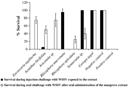
  • Under the first category of experiments, when shrimps were challenged with WSSV exposed to the extracts from R. mucronata, Sonneratia sp. and C. tagal significantly higher survival (95%, 100%, and 100% respectively) (Pb0.001) could be obtained.
  • Meanwhile, the batches of shrimps injected with WSSV exposed to the aqueous extracts of E. agallocha, Avicennia sp., and R. apiculata did not survive alike the positive control.
  • Meanwhile, comparatively lower survival was observed on feeding with the extracts from the other plants subsequent to WSSV challenge (75% with E. agallocha and Avicennia sp., 50% with A. ilicifolius, 40% with Sonneratia sp., 25% with R. apiculata, and no shrimp survived on feeding the extract of R. mucronata) (Pb0.001, Fig. 1).
  • On comparing the per cent survival of animals under the above two experimental conditions with the plant extracts (independent t-test), the one from C. tagal alone gave a uniformly higher rate of survival, the difference between the twowas least significant (PN0.05, Fig. 1).
  • With all other plant extracts significant variations between the two modes of experiments could be observed (Pb0.05).

3.2. Diagnostic PCR of the experimental animals

  • The virucidal property of the mangrove plant extracts and the protective effects of the samewere assessed by PCR ofWSSV extracted from the gill tissue of the experimental animals.
  • Meanwhile, the animals which were found dead after the administration of WSSV exposed to the extract of R. mucronata were PCR negative to WSSV.
  • The ones which survived the WSSV challenge after receiving the diet coated with the extracts from Sonneratia sp, Avicennia sp, E. agallocha, and A. ilicifolius were also negative to WSSV, however, the dead ones altogether were PCR positive.
  • The animals injected with the extract from positive control animals showed signs of WSSV and were PCR positive to the virus culminating in mortality (Table 1).

3.4. HPLC analysis and preliminary phytochemical investigation of crude aqueous extract from C. tagal

  • Repeated HPLC analysis of the crude aqueous extract generated uniformly a fingerprint of 25 peaks including 7 large well separated ones (Fig. 4).
  • Preliminary phytochemical investigation of the crude extract from C. tagal revealed the presence of appreciable quantities of polyphenols, saponins, sterols and flavonoids, and lesser quantities of alkaloids and cardiac glycosides.
  • The aqueous extract (500 ml) prepared from 50 g dried leaf of C. tagal yielded on average 3–4 g dry matter on lyophylization which contained the active fractions.
  • Based on a dye test conducted initially and the data on the survival of shrimp subsequent to challenge following administration of the extract along with diet, it could be realized that the binder, 4% aqueous gelatin, used in this study could effectively deliver the active fractions to shrimp.

4. Discussion

  • Extracts frommangrove plants and associates have been usedworld wide for medicinal purposes, and having been recorded around 349 metabolites it turns out to be a rich source of steroids, diterpenes and triterpenes, saponins,flavonoids, alkaloidsand tannins (Bandaranayake, 2002; Zhanget al., 2005; Pakhathirathienet al., 2005;Heet al., 2007;Wu et al., 2008).
  • In this experiment 74% survival of P. monodon wasobtained on administering the extract at 800 mg/kg bodyweight.
  • The virucidal property of the aqueous extracts of R. mucronata, Sonnaratia sp. and C. tagal when administered along with WSSV suspension at a 1:1 ratio after incubation for 3 h at room temperature suggested the presence of molecules in the preparation which could inactivate the virus.
  • The virucidal property of the aqueous extract of C. tagal was demonstrated through the total survival obtained on a challenge with the virus suspension exposed to the extract and the non infectivity of the tissue extracts of the ones that survived.
  • Pentacyclic triterpenes have been considered as a major bioactive group used as inhibitor of tumor cells, induction of apoptosis and also in antiviral therapy of AIDS (He et al., 2007).

Did you find this useful? Give us your feedback

Figures (5)
Citations
More filters
Journal ArticleDOI
TL;DR: The drug TP22C derived from Momordica charantia is a potent anti-white spot syndrome virus drug that showed 86% survivability in host.
Abstract: Objective To screen, isolate and optimize anti-white spot syndrome virus (WSSV) drug derived from various terrestrial plants and to evaluate the efficacy of the same in host–pathogen interaction model.

22 citations


Cites background or methods from "In vivo screening of mangrove plant..."

  • ...Previous researchers also used a viral titer of 1伊10 in their experiments[31]....

    [...]

  • ...monodon, researchers could obtain 100% survivability[31]....

    [...]

  • ...Antiviral activities of aqueous extracts from plants are well established and this includes reports on the antiviral activity of plant extracts against WSSV, too[29-31,39,50,53-60]....

    [...]

Journal ArticleDOI
TL;DR: The overall results suggest that the aqueous extract from C. tagal can protect P. monodon from white spot syndrome virus infection.
Abstract: White spot syndrome virus (WSSV), the most contagious pathogen of cultured shrimp, causes mass mortality, leading to huge economic loss to the shrimp industry. The lack of effective therapeutic or prophylactic measures has aggravated the situation, necessitating the development of antiviral agents. With this objective, the antiviral activity in the aqueous extract of a mangrove plant Ceriops tagal in Penaeus monodon was evaluated. The Ceriops tagal aqueous extract (CTAE) was non-toxic to shrimps at 50 mg/ml when injected intramuscularly at a dosage of 10 μL/animal (0.5 mg/animal) and showed a protective effect against WSSV at 30 mg/ml when mixed with WSSV suspension at a 1:1 ratio. When the extract was administered along with the diet and the animals were challenged orally, there was a dose-dependent increase in survival, culminating in 100 % survival at a concentration of 500 mg/kg body weight/day. Neither hypertrophied nuclei nor the viral envelope protein VP28 could be demonstrated in surviving shrimps using histology and indirect immunofluorescence histochemistry (IIFH), respectively. To elucidate the mode of action, the temporal expression of WSSV genes and shrimp immune genes, including antimicrobial peptides, was attempted. None of the viral genes were found to be expressed in shrimps that were fed with the extract and challenged or in those that were administered CTAE-exposed WSSV. The overall results suggest that the aqueous extract from C. tagal can protect P. monodon from white spot syndrome virus infection.

22 citations


Cites background from "In vivo screening of mangrove plant..."

  • ...A preliminary phytochemical study revealed that Ceriops tagal aqueous extract (CTAE) contains alkaloids, flavonoids, polyphenolics, cardiac glycosides, saponins and sterols, and an HPLC fingerprint was also generated [30]....

    [...]

Journal ArticleDOI
TL;DR: The data indicate that sodium alginate extracted from brown seaweed and fed to P. monodon can retard progression of WSSV disease.
Abstract: Sodium alginate extracted from brown seaweed Sargassum wightii (16.35 ± 1.42%, mean [±SD] yield from 5 extractions) was prepared as a powder or beads and used to enrich Artemia nauplii at concentrations of 100, 200, 300 and 400 mg l-1. The alginate-enriched nauplii were fed to Penaeus monodon shrimp postlarvae (PL) stage 15 (PL15, i.e. 15 d old) for 20 d. Mean weight gain and specific growth rate over this period were 0.24 g and 15.8%, respectively, in PL groups not fed alginate, and 0.20-0.28 g and 14.7-16.5%, respectively, in PL groups fed alginate. Amongst PL35 then challenged with white spot syndrome virus (WSSV) by immersion, all PL not fed alginate died within 9 d. However, amongst PL fed the 4 concentrations of alginate powder or beads, mortality rates reduced with increasing alginate concentration, and between 25 and 32% PL remained alive when the bioassay was terminated on Day 21. Amongst alginate-fed PL groups compared with the control group, mortality was reduced by 26.5 to 58.4%. Nested PCR detection of WSSV revealed sodium alginate concentration-dependent reductions in infection loads. The data indicate that sodium alginate extracted from brown seaweed and fed to P. monodon can retard progression of WSSV disease.

17 citations


Cites background from "In vivo screening of mangrove plant..."

  • ...More recently, Cereops tagal mangrove ex - tracts have been screened for anti-WSSV activity in P. monodon (Sudheer et al. 2011), and Litopenaeus vannamei immersed in seawater contai ning Sargassum hemiphyllum var. chinensis powder showed in - creased protection against Vibrio algino lyticus and…...

    [...]

Journal ArticleDOI
TL;DR: This review expresses in detail the versatile uses of phytotherapies and their based nanoformulation as a promising tool for disease management in aquaculture.

16 citations

Journal ArticleDOI
TL;DR: The overall results of the present study confirmed that acetone and petroleum ether extracts of P. amarus have significant antiviral effect against WSSV.
Abstract: Repeatedly the shrimp culturing industry faces serious economic loss due to white spot disease caused by white spot syndrome virus (WSSV) in penaeid shrimp. The present study was carried out to determine the antiviral activity of traditional Indian medicinal herb Phyllanthus amarus against WSSV in freshwater crab, Paratelphusa hydrodomous (Herbst). For experimental challenge, crabs were injected with three different solvent extracts of P. amarus (aqueous, acetone and petroleum ether) along with WSSV. The efficacy of the extracts was confirmed by bioassay, histopathology and reverse transcriptase – polymerase chain reaction (RT-PCR). The overall results of the present study confirmed that acetone and petroleum ether extracts of P. amarus have significant antiviral effect against WSSV.

15 citations

References
More filters
Journal ArticleDOI

17,196 citations


"In vivo screening of mangrove plant..." refers methods in this paper

  • ...Viral infectivity titre was determined as the extractable virus and expressed as LD 50 in shrimp following Reed and Muench (1938)....

    [...]

Book
09 Jan 2018
TL;DR: Indian medicinal plants/, Indian medicinal plants /, مرکز فناوری اطلاعات و اصاع رسانی, کδاوρزی
Abstract: Indian medicinal plants / , Indian medicinal plants / , مرکز فناوری اطلاعات و اطلاع رسانی کشاورزی

8,252 citations

Book
01 Jan 1956

5,524 citations


"In vivo screening of mangrove plant..." refers background in this paper

  • ...They have been used in folklore medicine for treatment of several diseases (Kirtikar and Basu, 1935; Chopra et al., 1956; Datta and Datta, 1982)....

    [...]

Book
01 Jan 1996

824 citations


"In vivo screening of mangrove plant..." refers background in this paper

  • ...…an enveloped non occluded DNA (300 kb) virus of the family Nimaviride under the new genus Wispovirus (Mayo, 2002), is the most devastating shrimp pathogen ever isolated and studied; it causes total mortality to a rearing stock within 3–7 days of infection in a culture system (Lightner, 1996)....

    [...]

  • ...White spot syndrome virus (WSSV), an enveloped non occluded DNA (300 kb) virus of the family Nimaviride under the new genus Wispovirus (Mayo, 2002), is the most devastating shrimp pathogen ever isolated and studied; it causes total mortality to a rearing stock within 3–7 days of infection in a culture system (Lightner, 1996)....

    [...]

Frequently Asked Questions (18)
Q1. What are the contributions mentioned in the paper "In vivo screening of mangrove plants for anti wssv activity in penaeus monodon, and evaluation of ceriops tagal as a potential source of antiviral molecules" ?

In this paper, the authors investigated the anti-White Spot Syndrome Virus ( WSSV ) activity in mangrove plants and found that WSSV can cause total mortality to a rearing stock within 3-7 days of infection in a culture system. 

As the aqueous extract from C. tagal alone could give protection to all animals tested against WSSV under the dual experimental conditions, C. tagal was identified for further studies. Further investigations are necessary in this direction. When shrimps were fed on the aqueous extract coated feed and subsequently challenged withWSSV, the viral DNA could not be detected in the tissue suggesting that the virus had neither invaded the host tissue nor multiplied. The virucidal property of the aqueous extracts of R. mucronata, Sonnaratia sp. and C. tagal when administered along with WSSV suspension at a 1:1 ratio after incubation for 3 h at room temperature suggested the presence of molecules in the preparation which could inactivate the virus. 

Pentacyclic triterpenes have been considered as a major bioactive group used as inhibitor of tumor cells, induction of apoptosis and also in antiviral therapy of AIDS (He et al., 2007). 

Diterpenes and triterpenes are the main natural products isolated from C. tagal (Zhang et al., 2005, Pakhathirathien et al., 2005, He et al., 2007). 

Cardiac glycosides have been demonstrated to have an inhibitory activity on themultiplication of herpex simplex virus (Dodson et al., 2007). 

On realizing the aqueous extract of C. tagal as the most effective preparation to protect shrimp from WSSV, it was subjected for HPLC analysis to generate HPLC fingerprint. 

Based on a dye test conducted initially and the data on the survival of shrimp subsequent to challenge following administration of the extract along with diet, it could be realized that the binder, 4% aqueous gelatin, used in this study could effectively deliver the active fractions to shrimp. 

The aqueous extract (500 ml) prepared from 50 g dried leaf of C. tagal yielded on average 3–4 g dry matter on lyophylization which contained the active fractions. 

The data generated on the survival of shrimp on administering the suspension of WSSV exposed to the plant extracts, and the data generated on the survival when challenged with WSSV subsequent to oral administration of the former were statistically analyzed employing the χ2 test. 

When shrimps were fed on the aqueous extract coated feed and subsequently challenged withWSSV, the viral DNA could not be detected in the tissue suggesting that the virus had neither invaded the host tissue nor multiplied. 

Confirmation of anti WSSV activityTo confirm the antiviral activity detected in the segregated plant species (C. tagal) intramuscular administration of virus suspension exposed to the plant extract, and oral administration of the plant extract and subsequent oral challenge were repeated in a batch of 24 animals and assayed by way of nested PCR. 

In their study, the quantity of the extract in the administered feed was nearly 1.0% of the total feed delivered at 500 mg/kg body weight per day. 

Balasubramanian et al. (2008) on feeding 2% aqueous extract of C. dactylon coated feed to P. moodon could obtain 100% survival and the survived animals were PCR negative. 

Viability of WSSV in suspension was checked by injecting 10 μl to a batch of apparently healthy shrimp (6 nos) and mortality confirmed over a period of 3 to 7 days. 

Under the second category of experiments, all the shrimps which were fed on the lyophilized aqueous extract of C. tagal could survive (100%) when orally challenged with WSSV (Pb0.001). 

The virucidal property of the aqueous extract of C. tagal was demonstrated through the total survival obtained on a challenge with the virus suspension exposed to the extract and the non infectivity of the tissue extracts of the ones that survived. 

Based on these evidences the authors conclude that the antiviral activity of the C. tagal aqueous extract might be due to one of the above compounds or due to their synergistic action. 

Confirmation of anti WSSV activity in aqueous extract of C. tagalSubsequently, the antiviral activity of the aqueous extract of C. tagal was reexamined by repeating both the experiments in a batch of 24 animals and on completion of the experiment after 15 days the animals were nested PCR negative, and when a tissue extract was passaged onto a fresh batch of animals none of them showed anyclinical signs of WSSV infection and remained negative to nested PCR.