scispace - formally typeset
Open AccessJournal ArticleDOI

Killing adherent and nonadherent cancer cells with the plasma pencil

Mounir Laroussi, +2 more
- 08 Jan 2015 - 
- Vol. 10, Iss: 2, pp 029401
Reads0
Chats0
TLDR
Results indicate that plasma consistently shows a delayed killing effect that is dose dependent, and there is some evidence that apoptosis is one of the pathways that leads to the death of the cells, indicating that plasma initiates cell signaling pathways.
Abstract
The application of low temperature plasmas in biology and medicine may lead to a paradigm shift in the way various diseases can be treated without serious side effects. Low temperature plasmas generated in gas mixtures that contain oxygen or air produce several chemically reactive species that have important biological implications when they interact with eukaryotic or prokaryotic cells. Here, a review of the effects of low temperature plasma generated by the plasma pencil on different cancerous cells is presented. Results indicate that plasma consistently shows a delayed killing effect that is dose dependent. In addition, there is some evidence that apoptosis is one of the pathways that leads to the death of the cells, indicating that plasma initiates cell signaling pathways.

read more

Content maybe subject to copyright    Report

Old Dominion University
ODU Digital Commons
/)'64-'%/20376)41+-1))4-1+%'7/69
7&/-'%6-215
/)'64-'%/20376)41+-1))4-1+

Killing Adherent and Nonadherent Cancer Cells
with the Plasma Pencil
Mounir Laroussi
Old Dominion University0/%427552(7)(7
Soheila Mohades
Old Dominion University
Nazir Barekzi
Old Dominion University
2//286,-5%1(%((-6-21%/824.5%6 ,<35(-+-6%/'2002152(7)(7)')$*%'$37&5
%462*6,) -20)(-'%/1+-1))4-1+%1(-2)1+-1))4-1+200215 )23/%505200215%1(
6,) /%50%%1()%0 ,95-'5200215
;-546-'/)-5&427+,662927*24*4))%1(23)1%'')55&96,)/)'64-'%/20376)41+-1))4-1+%6#-+-6%/2002156,%5&))1%'')36)(*24
-1'/75-21-1/)'64-'%/20376)41+-1))4-1+%'7/69 7&/-'%6-215&9%1%76,24-:)(%(0-1-564%6242*#-+-6%/20021524024)-1*240%6-21
3/)%5)'216%'6 (-+-6%/'2002152(7)(7
!)325-6249-6%6-21
%42755-271-42,%()5"2,)-/%%1(%4).:-%:-4-//-1+(,)4)16%1(21%(,)4)16%1')4)//58-6,6,) /%50% )1'-/
 Electrical & Computer Engineering Faculty Publications
,<35(-+-6%/'2002152(7)(7)')$*%'$37&5
4-+-1%/ 7&/-'%6-21-6%6-21
%42755-2,%()5"%4).:--//-1+%(,)4)16%1(121%(,)4)16'%1')4')//58-6,6,)3/%50%3)1'-/ Biointerphases,
1033(2-

Killing adherent and nonadherent cancer cells with the plasma pencil
Mounir Laroussi, Soheila Mohades, and Nazir Barekzi
Citation: Biointerphases 10, 029401 (2015); doi: 10.1116/1.4905666
View online: http://dx.doi.org/10.1116/1.4905666
View Table of Contents: http://scitation.aip.org/content/avs/journal/bip/10/2?ver=pdfcov
Published by the AVS: Science & Technology of Materials, Interfaces, and Processing
Articles you may be interested in
Biomechanical investigation of colorectal cancer cells
Appl. Phys. Lett. 105, 123701 (2014); 10.1063/1.4896161
Cell-matrix adhesion characterization using multiple shear stress zones in single stepwise microchannel
Appl. Phys. Lett. 105, 083701 (2014); 10.1063/1.4892666
Selective cell capture and analysis using shallow antibody-coated microchannels
Biomicrofluidics 6, 044117 (2012); 10.1063/1.4771968
Selective killing of ovarian cancer cells through induction of apoptosis by nonequilibrium atmospheric pressure
plasma
Appl. Phys. Lett. 100, 113702 (2012); 10.1063/1.3694928
Morphological observation and adhesive property measurement on human ovary carcinoma cells by atomic force
microscopy
J. Vac. Sci. Technol. B 27, 1370 (2009); 10.1116/1.3066057

Killing adherent and nonadherent cancer cells with the plasma pencil
Mounir Laroussi,
a)
Soheila Mohades, and Nazir Barekzi
Plasma Engineering & Medicine Institute, Old Dominion University, Norfolk, Virginia 23529
(Received 25 November 2014; accepted 29 December 2014; published 8 January 2015)
The application of low temperature plasmas in biology and medicine may lead to a paradigm
shift in the way various diseases can be treated without serious side effects. Low temperature
plasmas generated in gas mixtures that contain oxygen or air produce several chemically
reactive species that have important biological implications when they interact with eukaryotic
or prokaryotic cells. Here, a review of the effects of low temperature plasma generated by
the plasma pencil on different cancerous cells is presented. Results indicate that plasma
consistently shows a delayed killing effect that is dose dependent. In addition, there is
some evidence that apoptosis is one of the pathways that leads to the death of the cells,
indicating that plasma initia tes cell signaling pathways.
V
C
2015 American Vacuum Society.
[http://dx.doi.org/10.1116/1.4905666]
I. INTRODUCTION
Plasma Medicine is a field of research that deals with
the biomedical applications of low temperature plasmas
(LTPs) and m ost specifically plasmas generated at atmos-
pheric pressure and at biologically tolerable tempera-
tures.
1,2
These types of plasmas can be safely applied to
biological systems (cells, t issues, organs, etc.) without
causing thermal damage. The plasma can be tuned to
achieve various effects ranging from killing pathogens, to
stimulating the proliferation of healthy cells, to destroying
cancer cells.
13
Tuning the plasma t o achieve desired
effects involves judicious sele ction of the operating gas
mixture, the type of waveform of the applied voltage (DC,
pulsed DC, RF, pulsed RF, microwave, etc.), and careful
adjustment of the amount of applied power to influence the
electrons energy distribution function and to control the
fluxes of the reactive species. There is a general agree-
ment, based on overwhelming experimental evidence, that
the plasma-generated reactive oxygen species (ROS) and
reactive nitrogen species (RNS) play significant roles in
most of the observed effects of plasma on biological cells
and biological systems. This comes as no surprise as it is
well known from cell biology investigations that ROS and
RNS are involved in various biochemical events that occur
on the cellular and subcellular levels. Some of these
include lipids peroxidation, DNA damage, and the trigger-
ing of cellular signals.
14
Of all the possible applications of LTP in medical therapy
none is expected to have as much of an important scientific
and societal impact as cancer treatment. Today, the work on
cancer is becoming the most exciting topic in plasma medi-
cine research. Here, first what is known about the mecha-
nisms of action of LTP against cancer cells is briefly
reviewed, then the results of the application of a particular
LTP source termed the “plasma pencil” to kill cancer cells
are presented.
II. MECHANISMS OF ACTION OF LTP AGAINST
CANCER CELLS
In general, LTP is applied to cells that are either sub-
merged in a liquid medium or covered by a thin layer of a
liquid/fluid. Therefore, the reactive species generated by the
plasma in the gaseous state have to go through a gas–liquid
interface and then diffuse into the bulk of the liquid. Since
most plasma-generated reactive species are short-lived
whereas the plasma-induced biological effects on cells have
been observed only a relatively long time later, we hypothe-
size that the short lived species first react with the medium
to create reaction by-products that are long-lived and it is
these species that subsequently interact with the cells. These
longer-lived reaction byproducts would include H
2
O
2
,
NO
2
,NO
3
,RO
2
(organic peroxide), etc. Our work showed
that if a medium is treated by LTP, then cells are seeded into
this plasma activated medium (PAM), the effects on the cells
greatly depends on the “age” of the PAM.
5
By age it is
meant how long a time PAM is stored before applied on the
cells. The older the PAM, the lower the killing effect on
cells. Fresh PAM (not aged/applied right after plasma activa-
tion) has the strongest killing effect. Hence, we concluded
that plasma reaction byproducts in the medium are responsi-
ble for killing the cells, but as their concentrations decrease
with PAM storage/aging time (hours) most cells would be
able to cope and survive. It is important to note that the kill-
ing of cells by PAM occurs in a delayed manner, the same as
for the case when cells are directly exposed to LTP.
Based on experimental evidence, several investigators
reported that cancer cells appear to be more vulnerable
to LTP than their healthy counterparts.
6,7
Keidar and co-
workers reported that LTP causes an increase in the expres-
sion of the oxidative stress reporter cH2A.X (pSer 139) and
a decrease in DNA replication in the S-phase of the cell
cycle.
8
The S-phase is the phase in the cell cycle when DNA
replication occurs. These effects were much less pronounced
in the healthy cells. Since LTP appears to interfere with the
DNA replication phase and since cancer cells multiply more
rapidly than their healthy counterparts, they are much more
a)
Electronic mail: mlarouss@odu.edu
029401-1 Biointerphases 10(2), June 2015 1934-8630/2015/10(2)/029401/7/$30.00
V
C
2015 American Vacuum Society 029401-1

susceptible to LTP-induced damage. In general, cancer cells
have much higher metabolic rate than healthy cells and
therefore exhibit higher levels of intracellular concentrations
of ROS and RNS. Hence, exposure of cancer cells to LTP
leads to an even higher intracellular oxidative stress, there-
fore causing their death.
Other investigators have reported that cell signaling plays
an important role in the interaction of LTP with cancer cells.
One such signaling pathway is the activation of caspases.
Caspases are proteins that play a crucial role in apoptosis or
programmed cell death. The lack of initiation of apoptosis
leads to uncontrolled cell multiplication and tumor
development.
Laroussi and co-workers studied caspase-3 activation in
the case of squamous cell carcinoma and found higher level
of caspase-3 activation in cells treated by LTP than in con-
trol samples (untreated cells), indicating that LTP induces
apoptosis in these cells.
9
Ishaq et al. suggested that because
tumor cells are defective in several regulatory signaling
pathways they exhibit metabolic imbalance, which leads to a
lack of cell growth regulation.
10
According to these authors,
LTP-generated ROS affect the metabolism of the cancer
cells by impairing redox balance, which leads to slowing
down or arresting the proliferation of the cells. These inves-
tigators also reported that LTP up-regulates intracellular
ROS levels and induce apoptosis in melanoma.
11
They iden-
tified that LTP exposure causes a differential expression of
tumor necrosis factor (TNF) family members in the cancer-
ous cells but not in the normal cells. They found that apopto-
sis in the cancer cells was induced by apoptosis signal kinase
1 (ASK1), which is activated by TNF signaling.
III. PLASMA PENCIL
The application of LTP in biology and medicine relies on
the availability of various sources that can produce plasmas
with gas temperatures less than about 40
C, and at atmos-
pheric pressure (760 Torr). This is crucial since if LTP is
to be used as a basis for a medical therapy it has to be gener-
ated at surrounding room conditions such as in a hospital or
clinic setting. Among the various LTP sources, low tempera-
ture, atmospheric pressure plasma jets are most practical for
biomedical applications because they readily provide plas-
mas to a target at room conditions.
1214
These jets can be
powered by sources covering a wide frequency spectrum
that ranges from DC to microwaves. Various types of plasma
jets have therefore been developed with designs tailored for
specific needs. One such device, the plasma pencil, a hand-
held plasma jet generator developed in our laboratory, is pre-
sented here in some detail.
The electrodes of the plasma pencil are disk-shaped and
spaced by a gap that can be varied from 0.5 to1 cm (see Fig.
1).
15
Each of the two electrodes is made of a thin copper ring
attached to the surface of a centrally perforated dielectric
disk. To ignite the plasma, high voltage (few kV) pulses at
repetition rates in the 1–10 kHz range are applied between
the two electrodes and a gas mixture (such as helium and ox-
ygen) is flown through the holes of the electrodes (flow rates
in the 1–10 l/min range). Plasma plumes reaching lengths up
to 5 cm can be launched through the hole of the outer elec-
trode and in the surrounding room air.
15
The length of the
plume depends on the gas flow rate, the magnitude of the
applied voltage pulses, their widths, and the frequency. The
plasma plume exhibits low temperature and can be touched
by bare hands without causing any harm. A photograph of
the plume emitted by the plasma pencil and touching the
author’s hand is shown in Fig. 2. Since the plasma plume
does not cause thermal damage even to delicate tissue, it can
be applied to disinfect skin wounds resulting from cuts or
burns and to destroy cancer cells without damaging healthy
cells.
Using a combination of diagnostics involving intensified
charge coupled device imaging, optical emission spectros-
copy, and electrical probes it was discovered that the plasma
plume is in fact not a continuous volume of plasma, as they
appear to the naked eye, but discrete small volumes of
plasma referred to as “plasma bullets,” which travel at very
high velocities.
16,17
Unlike regular plasma streamers, which
appear at random times and spaces, these plasma bullets ex-
hibit behavior that is highly repeatable and predictable. Lu
FIG. 1. Schematic of the plasma pencil.
029401-2 Laroussi, Mohades, and Barekzi: Killing adherent and nonadherent cancer cells 029401-2
Biointerphases, Vol. 10, No. 2, June 2015

and Laroussi first proposed photoionization as a necessary
mechanism to explain the propagation of the plasma bul-
lets;
17
however, today there is consensus that plasma bullets
are “guided” ionization waves where the electric field at the
head of the plasma bullet plays an important role in its prop-
agation characte ristics.
14
Because plasma bullets are launched in air, they produce
very interesting reactive chemistry that can be exploited in
biological and medical applications. Reactive oxygen spe-
cies, such as O, OH, O
2
, an d reac tive nitrogen species,
such as NO, NO
2
, which are known to have biological
implications, are abundantly generated by the plasma
bullets.
IV. EFFECTS OF THE PLASMA PENCIL ON
CANCER CELLS
To investigate the efficacy of the plasma pencil against
cancer both adherent and nonadherent cancerous cells
were tested. As a representative of nonadherent cell lines
leukemia cells were selected. As a dherent cells, squamous
cell carcinoma and prostate cancer cells were used. The
following is a review of the results obtained. The experi-
mental setup used in these studies is shown in Fig. 3.The
reader is hereby notified that the following is not a
review of the field but rather a review of our own results.
These results were originally published elsewhere, in var-
ious scientific journals. References to the original papers
are provided, where more details of the experiments can
be found. The remainder of this paper mainly summarizes
our work of the last few years on cancer application
using our low temperature plasma generator, the plasma
pencil.
A. Case of nonadherent cells
In this study, T-cell line (ATCC No. CCL-119; aka
CCRF-CEM) originally isolated from the blood of a patient
with acute lymphoblastic leukemia was used. The CCRF-
CEM cells are nonadherent leukemia cancer cells, obtained
from the American Type Culture Collection. They were
grown in complete growth media in 75 cm
2
vented sterile
polystyrene tissue culture flasks at 37
C in a humidified
atmosphere containing 5% CO
2
. The growth media consisted
of buffered RPMI-1640 media supplemented with 10% fetal
bovine serum, 1% antibiotics (penicillin/streptomycin), and
1% glutamine.
Initial experiments revealed that plasma kills the leu-
kemia cells and that the effects of a single dose of
plasma continue for up to few days. Figures 4(a) and
4(b) illustrate results using CCRF-CEM leukemia cells,
in vitro, treated at various doses and evaluated at 12
and 36 h post-treatment, respectively.
18
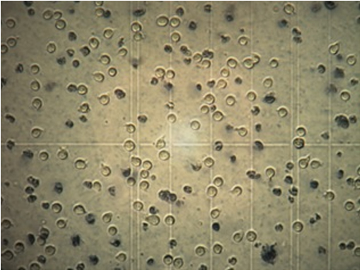
Figure 5 shows
images that illustrate t he difference in color between
cells that took up the trypan blue dye (dead) and those
that did not (live).
The results revealed that the cell viability did not differ
dramatically at time 0 h postplasma treatment even at the
highest dose of treatment which was a plasma exposure of
10 min. However, when post-treatment analysis of cell via-
bility was determined at 12, 36, or 60 h, statistically signifi-
cant killing was observed even at the lower plasma doses.
Cell survivability data analyzed at 12, 36, or 60 h postplasma
treatment revealed that there was a threshold treatment time
of 3 min beyond which a highly statistically significant
increase in leukemia cell death was obtained.
18
The results
from this study indicate that there is a dose dependent
response in the induction of cell death of leukemia cells and
single doses of plasma treatment continue killing cells up to
2.5 days post treatment. The delayed response of cell death
may be attributed to cell signaling cas cades that may result
in apoptosis. However, for higher doses, such as for 10 min
exposure, necrotic cells were observed immediately after
plasma exposure.
FIG. 3. Experimental setup for the exposure of samples to the plasma pencil.
FIG. 2. Cold plasma plume touching the author’s hand.
029401-3 Laroussi, Mohades, and Barekzi: Killing adherent and nonadherent cancer cells 029401-3
Biointerphases, Vol. 10, No. 2, June 2015

Figures
Citations
More filters
Journal ArticleDOI

Reactive species in non-equilibrium atmospheric-pressure plasmas: Generation, transport, and biological effects

TL;DR: In this paper, the most important mechanisms of generation and transport of the key species in the plasmas of atmospheric-pressure plasma jets and other non-equilibrium atmospheric pressure plasms are introduced and examined from the viewpoint of their applications in plasma hygiene and medicine and other relevant fields.
Journal ArticleDOI

Cold atmospheric plasma, a novel promising anti-cancer treatment modality.

TL;DR: This review provides a comprehensive introduction of the basics of CAP, state of the art research in this field, the primary challenges, and future directions to cancer biologists.
Journal ArticleDOI

Perspective: The physics, diagnostics, and applications of atmospheric pressure low temperature plasma sources used in plasma medicine

TL;DR: In this article, the authors used low temperature plasma discharges as sources of chemically reactive species that can be transported to interact with biological media, cells, and tissues and induce impactful biological effects.
Journal ArticleDOI

Plasma Medicine: A Brief Introduction

TL;DR: This mini review is to introduce the readers of Plasma to the field of plasma medicine, a multidisciplinary field of research at the intersection of physics, engineering, biology and medicine.
Journal ArticleDOI

Cold Plasma in Medicine and Healthcare: The New Frontier in Low Temperature Plasma Applications

TL;DR: A mini review of the physical and biochemical mechanisms whereby low temperature plasma affects biological cells on macroscopic and microscopic scales can be found in this article, where a thorough understanding of these mechanisms is bound to lead to the development of novel plasma-based medical therapies.
References
More filters
Journal Article

Atmospheric gas plasma–induced ROS production activates TNF-ASK1 pathway for the induction of melanoma cancer cell apoptosis

TL;DR: Atmospheric gas plasmas up-regulate intracellular ROS levels and induce apoptosis in melanoma cells and evidence for TNF-signaling dependence of ASK1-mediated apoptosis suggests possible mechanisms for AGP activation and regulation of apoptosis-signalling pathways in tumor cells.
Journal ArticleDOI

Dose-dependent killing of leukemia cells by low-temperature plasma

TL;DR: The outcome of this study revealed that the effect of plasma exposure was not immediate, but had a delayed effect and increasing the time of plasma Exposure resulted in increased leukemia cell death.
Journal ArticleDOI

Effects of Low Temperature Plasmas on Cancer Cells

TL;DR: Conventional cancer treatments, some of the plasma devices that are currently used and the influence of BTPs on cancer cells such as melanomas, carcinomas, and leukemia are reviewed and discussed.
Journal ArticleDOI

Efficacy of Low Temperature Plasma against SCaBER Cancer Cells

TL;DR: Caspase-3 assays reveal that plasma can activate the cells' apoptotic pathways and alter cell morphology, cell reattachment, and kill SCaBER cells in an exposure time dependent and delayed manner.
Related Papers (5)
Frequently Asked Questions (18)
Q1. What contributions have the authors mentioned in the paper "Killing adherent and nonadherent cancer cells with the plasma pencil" ?

This Article is brought to you for free and open access by the Electrical & Computer Engineering at ODU Digital Commons. 

The length of the plume depends on the gas flow rate, the magnitude of the applied voltage pulses, their widths, and the frequency. 

Caspase-3 is a protein involved in the apoptotic pathway that interacts with caspase-8 and caspase-9 through a sequential process that activates cell apoptosis. 

14Because plasma bullets are launched in air, they produce very interesting reactive chemistry that can be exploited in biological and medical applications. 

Reactive oxygen species, such as O, OH, O2 , and reactive nitrogen species, such as NO, NO2, which are known to have biological implications, are abundantly generated by the plasma bullets. 

In general, cancer cells have much higher metabolic rate than healthy cells and therefore exhibit higher levels of intracellular concentrations of ROS and RNS. 

6,7 Keidar and coworkers reported that LTP causes an increase in the expression of the oxidative stress reporter cH2A.X (pSer 139) and a decrease in DNA replication in the S-phase of the cell cycle. 

Plasma plumes reaching lengths up to 5 cm can be launched through the hole of the outer electrode and in the surrounding room air. 

They were grown in complete growth media in a vented tissue culture flask and were incubated at 37 C in a humidified atmosphere containing 5% CO2. 

For 5 min plasma treatment, only about 25% of cells remained viable at 24 h postplasma exposure, indicating that the higher the dose of the plasma the lower the number of the cells that survived. 

18 The results from this study indicate that there is a dose dependent response in the induction of cell death of leukemia cells and single doses of plasma treatment continue killing cells up to 2.5 days post treatment. 

Initial experiments revealed that plasma kills the leukemia cells and that the effects of a single dose of plasma continue for up to few days. 

In this study, T-cell line (ATCC No. CCL-119; aka CCRF-CEM) originally isolated from the blood of a patient with acute lymphoblastic leukemia was used. 

Laroussi and co-workers studied caspase-3 activation in the case of squamous cell carcinoma and found higher level of caspase-3 activation in cells treated by LTP than in control samples (untreated cells), indicating that LTP induces apoptosis in these cells.9 Ishaq et al. suggested that because tumor cells are defective in several regulatory signaling pathways they exhibit metabolic imbalance, which leads to a lack of cell growth regulation. 

the reactive species generated by the plasma in the gaseous state have to go through a gas–liquid interface and then diffuse into the bulk of the liquid. 

starting with 24 h post-treatment, a slow but steady trend of cell death for plasma exposure times longer than 5 min was observed. 

The results revealed that the cell viability did not differ dramatically at time 0 h postplasma treatment even at the highest dose of treatment which was a plasma exposure of 10 min. 

The counts immediately after plasma pencil treatment at time 0 h029401-5 Laroussi, Mohades, and Barekzi: Killing adherent and nonadherent cancer cells 029401-5Biointerphases, Vol. 10, No. 2, June 2015reveal no dead cells, suggesting that there were no immediate physical effects.