scispace - formally typeset
Search or ask a question
Journal ArticleDOI

Preoperative Chemoradiotherapy for Esophageal or Junctional Cancer

31 May 2012-The New England Journal of Medicine (MASSACHUSETTS MEDICAL SOC)-Vol. 366, Iss: 22, pp 2074-2084
TL;DR: Preoperative chemoradiotherapy improved survival among patients with potentially curable esophageal or esophagogastric-junction cancer and the regimen was associated with acceptable adverse-event rates.
Abstract: A B S T R AC T BACKGROUND The role of neoadjuvant chemoradiotherapy in the treatment of patients with esophageal or esophagogastric-junction cancer is not well established. We compared chemoradiotherapy followed by surgery with surgery alone in this patient population. METHODS We randomly assigned patients with resectable tumors to receive surgery alone or weekly administration of carboplatin (doses titrated to achieve an area under the curve of 2 mg per milliliter per minute) and paclitaxel (50 mg per square meter of body-surface area) for 5 weeks and concurrent radiotherapy (41.4 Gy in 23 fractions, 5 days per week), followed by surgery. RESULTS From March 2004 through December 2008, we enrolled 368 patients, 366 of whom were included in the analysis: 275 (75%) had adenocarcinoma, 84 (23%) had squamous-cell carcinoma, and 7 (2%) had large-cell undifferentiated carcinoma. Of the 366 patients, 178 were randomly assigned to chemoradiotherapy followed by surgery, and 188 to surgery alone. The most common major hematologic toxic effects in the chemoradiotherapy–surgery group were leukopenia (6%) and neutropenia (2%); the most common major nonhematologic toxic effects were anorexia (5%) and fatigue (3%). Complete resection with no tumor within 1 mm of the resection margins (R0) was achieved in 92% of patients in the chemoradiotherapy–surgery group versus 69% in the surgery group (P<0.001). A pathological complete response was achieved in 47 of 161 patients (29%) who underwent resection after chemoradiotherapy. Postoperative complications were similar in the two treatment groups, and in-hospital mortality was 4% in both. Median overall survival was 49.4 months in the chemoradiotherapy– surgery group versus 24.0 months in the surgery group. Overall survival was significantly better in the chemoradiotherapy–surgery group (hazard ratio, 0.657; 95% confidence interval, 0.495 to 0.871; P = 0.003). CONCLUSIONS Preoperative chemoradiotherapy improved survival among patients with potentially curable esophageal or esophagogastric-junction cancer. The regimen was associated with acceptable adverse-event rates. (Funded by the Dutch Cancer Foundation [KWF Kankerbestrijding]; Netherlands Trial Register number, NTR487.)

Content maybe subject to copyright    Report

original article
The
new engl a nd jour nal
o f
medicine
n engl j med 366;22 nejm.org may 31, 2012
2074
Preoperative Chemoradiotherapy
for Esophageal or Junctional Cancer
P. van Hagen, M.C.C.M. Hulshof, J.J.B. van Lanschot, E.W. Steyerberg,
M.I. van Berge Henegouwen, B.P.L. Wijnhoven, D.J. Richel,
G.A.P. Nieuwenhuijzen, G.A.P. Hospers, J.J. Bonenkamp, M.A. Cuesta,
R.J.B. Blaisse, O.R.C. Busch, F.J.W. ten Kate, G.-J. Creemers, C.J.A. Punt,
J.T.M. Plukker, H.M.W. Verheul, E.J. Spillenaar Bilgen, H. van Dekken,
M.J.C. van der Sangen, T. Rozema, K. Biermann, J.C. Beukema,
A.H.M. Piet, C.M. van Rij, J.G. Reinders, H.W. Tilanus,
and A. van der Gaast, for the CROSS Group*
The authors’ full names, degrees, and affili-
ations are listed in the Appendix. Address
reprint requests to Dr. van der Gaast at
the Department of Medical Oncology,
Erasmus University Medical Center/Daniel
den Hoed Cancer Center, P.O. Box 2040,
3000 CA Rotterdam, the Netherlands, or at
a.vandergaast@erasmusmc.nl.
* The members of the Chemoradiothera-
py for Oesophageal Cancer Followed by
Surgery Study (CROSS) Group are list-
ed in the Supplementary Appendix,
available at NEJM.org.
N Engl J Med 2012;366:2074-84.
Copyright © 2012 Massachusetts Medical Society.
ABSTR ACT
BACKGROUND
The role of neoadjuvant chemoradiotherapy in the treatment of patients with esopha-
geal or esophagogastric-junction cancer is not well established. We compared chemo-
radiotherapy followed by surgery with surgery alone in this patient population.
METHODS
We randomly assigned patients with resectable tumors to receive surgery alone or
weekly administration of carboplatin (doses titrated to achieve an area under the
curve of 2 mg per milliliter per minute) and paclitaxel (50 mg per square meter of
body-surface area) for 5 weeks and concurrent radiotherapy (41.4 Gy in 23 fractions,
5 days per week), followed by surgery.
RESULTS
From March 2004 through December 2008, we enrolled 368 patients, 366 of whom
were included in the analysis: 275 (75%) had adenocarcinoma, 84 (23%) had squa-
mous-cell carcinoma, and 7 (2%) had large-cell undifferentiated carcinoma. Of the
366 patients, 178 were randomly assigned to chemoradiotherapy followed by surgery,
and 188 to surgery alone. The most common major hematologic toxic effects in the
chemoradiotherapy–surgery group were leukopenia (6%) and neutropenia (2%); the
most common major nonhematologic toxic effects were anorexia (5%) and fatigue (3%).
Complete resection with no tumor within 1 mm of the resection margins (R0) was
achieved in 92% of patients in the chemoradiotherapy–surgery group versus 69% in
the surgery group (P<0.001). A pathological complete response was achieved in 47 of
161 patients (29%) who underwent resection after chemoradiotherapy. Postoperative
complications were similar in the two treatment groups, and in-hospital mortality was
4% in both. Median overall survival was 49.4 months in the chemoradiotherapy–
surgery group versus 24.0 months in the surgery group. Overall survival was sig-
nificantly better in the chemoradiotherapy–surgery group (hazard ratio, 0.657; 95%
confidence interval, 0.495 to 0.871; P = 0.003).
CONCLUSIONS
Preoperative chemoradiotherapy improved survival among patients with potentially
curable esophageal or esophagogastric-junction cancer. The regimen was associated
with acceptable adverse-event rates. (Funded by the Dutch Cancer Foundation [KWF
Kankerbestrijding]; Netherlands Trial Register number, NTR487.)
The New England Journal of Medicine
Downloaded from nejm.org at VRIJE UNIVERSITEIT on October 29, 2013. For personal use only. No other uses without permission.
Copyright © 2012 Massachusetts Medical Society. All rights reserved.

Preoperative Chemoradiotherapy for Esophageal Cancer
n engl j med 366;22 nejm.org may 31, 2012
2075
W
ith new diagnoses in more than
480,000 patients annually, esophageal
cancer is the eighth most common can-
cer worldwide.
1
It is a highly lethal disease, causing
more than 400,000 deaths per year.
2
The incidence
of esophageal adenocarcinoma is rapidly rising,
whereas that of squamous-cell carcinoma remains
unchanged.
3
Despite adequate preoperative stag-
ing, 25% of patients treated with primary sur-
gery have microscopically positive resection mar-
gins (R1), and the 5-year survival rate rarely
exceeds 40%.
4
The role of neoadjuvant chemoradiotherapy has
been debated for several decades. In most random-
ized trials, no survival benefit could be shown, and
the trials were criticized for inadequate trial de-
sign, samples that were too small, and poor out-
comes in the surgery-alone group. Meta-analyses
suggest a survival benefit from neoadjuvant chemo-
radiotherapy, albeit frequently at the cost of in-
creased postoperative morbidity and mortality.
5,6
We previously reported a phase 2 trial of neo-
adjuvant chemoradiotherapy consisting of weekly
administration of carboplatin and paclitaxel with
concurrent radiotherapy.
7
This regimen was as-
sociated with a low rate of serious toxic effects,
and a complete resection with no tumor within
1 mm of the resection margins (R0) was achieved
in all patients who underwent resection. These re-
sults encouraged us to initiate a multicenter, ran-
domized, controlled, phase 3 study comparing
neoadjuvant chemoradiotherapy followed by sur-
gery with surgery alone in patients with poten-
tially curable esophageal or esophagogastric-
junction carcinoma.
8
METHODS
ELIGIBILITY CRITERIA
Patients with histologically confirmed, potentially
curable squamous-cell carcinoma, adenocarci-
noma, or large-cell undifferentiated carcinoma of
the esophagus or esophagogastric junction (i.e.,
tumors involving both the cardia and the esopha-
gus on endoscopy) were eligible for inclusion in
the study. The upper border of the tumor had to be
at least 3 cm below the upper esophageal sphincter.
Patients who had proximal gastric tumors with
minimal invasion of the esophagus were excluded.
The length and width of the tumor could not ex-
ceed 8 cm and 5 cm, respectively. Only patients
with tumors of clinical stage T1N1 or T2-3N0-1
and no clinical evidence of metastatic spread
(M0), according to the International Union
against Cancer (UICC) tumor–node–metastasis
(TNM) classification,
9
were enrolled. Eligible pa-
tients were 18 to 75 years of age, had a World
Health Organization (WHO) performance status
score of 2 or lower (on a scale of 0 to 5, with 0
indicating fully active, 1 unable to carry out
heavy physical work, and 2 up and about more
than half the day but unable to work), and had
lost 10% or less of body weight. Patients also had
to have adequate hematologic, renal, hepatic, and
pulmonary function, as well as no history of other
cancer or previous radiotherapy or chemotherapy.
All patients provided written informed consent.
The institutional review board at each participating
center approved the study protocol.
8
The protocol,
including the statistical analysis plan, is available
with the full text of this article at NEJM.org. No
commercial support was involved in the study;
the drugs were purchased. No one who is not an
author contributed to the manuscript. The first,
fourth, and last authors vouch for the accuracy
and completeness of the reported data and the
fidelity of the study to the protocol.
STAGING
All patients underwent pretreatment staging. This
included a history taking; physical examination;
pulmonary-function tests, routine hematologic
and biochemical tests; upper gastrointestinal en-
doscopy with histologic biopsy and endoscopic
ultrasonography; computed tomography of the
neck, chest, and upper abdomen; and external ul-
trasonography of the neck, with fine-needle aspi-
ration of lymph nodes when cancer was suspected.
For the final analysis, the available endoscopic re-
ports were centrally reviewed.
TREATMENT
Chemotherapy
On days 1, 8, 15, 22, and 29, carboplatin targeted
at an area under the curve of 2 mg per milliliter
per minute and paclitaxel at a dose of 50 mg per
square meter of body-surface area were adminis-
tered intravenously. All patients were intravenously
premedicated with dexamethasone, clemastine,
and ranitidine as well as standard antiemetic
agents. The patients were closely monitored for
toxic effects of chemotherapy with the use of the
National Cancer Institute’s Common Terminolo-
gy Criteria for Adverse Events, version 3.0.
10
The New England Journal of Medicine
Downloaded from nejm.org at VRIJE UNIVERSITEIT on October 29, 2013. For personal use only. No other uses without permission.
Copyright © 2012 Massachusetts Medical Society. All rights reserved.

The
ne w engl and jour nal
o f
medicine
n engl j med 366;22 nejm.org may 31, 2012
2076
Radiotherapy
A total radiation dose of 41.4 Gy was given in 23
fractions of 1.8 Gy each, with 5 fractions admin-
istered per week, starting on the first day of the
first chemotherapy cycle. All patients were treated
by means of external-beam radiation. A detailed
description of the methods of administration of
chemotherapy and radiotherapy can be found in
Appendix 1 in the Supplementary Appendix, avail-
able at NEJM.org.
Surgery
Patients in the chemoradiotherapy–surgery group
underwent surgery as soon as possible after com-
pletion of chemoradiotherapy (preferably, within
4 to 6 weeks), and patients in the surgery group
were treated as soon as possible after randomiza-
tion. A transthoracic approach with two-field
lymph-node dissection was performed for tumors
extending proximally to the tracheal bifurcation.
For tumors involving the esophagogastric junction,
a transhiatal resection was preferred. Peritruncal
dissection was carried out with both approaches.
For all other tumors, the approach depended on the
characteristics of the patient and on local prefer-
ences. Gastric-tube reconstruction with a cervical
anastomosis was the preferred technique for re-
storing the continuity of the digestive tract.
PATHOLOGICAL ANALYSIS
Reports on pathological examination had to de-
scribe the tumor type and extension, lymph nodes,
and resection margins. In the absence of macro-
scopic tumor, any abnormal-appearing tissue
was paraffin-embedded in total in order to make
an adequate assessment for the presence of re-
sidual tumor and the effects of therapy.
To grade the response to therapy, we classified
the degree of histomorphologic regression into
four categories as follows: grade 1, no evidence of
vital residual tumor cells (pathological complete
response); grade 2, less than 10% vital residual
tumor cells; grade 3, 10 to 50%; and grade 4,
more than 50%.
11,12
If a vital tumor was present
at 1 mm or less from the proximal, distal, or cir-
cumferential resection margin, it was considered
to be microscopically positive (R1).
FOLLOW-UP
During the first year after treatment was com-
pleted, patients were seen every 3 months. In the
second year, follow-up took place every 6 months,
and then at the end of each year until 5 years af-
ter treatment. Late toxic effects, disease recur-
rence, and death were documented. Recurrences
were scored at the moment of the first recur-
rence. During follow-up, diagnostic investiga-
tions were performed only when recurrence was
suspected.
STATISTICAL ANALYSIS
We calculated that 175 patients were needed in
each group in order to detect a difference in me-
dian overall survival of 22 months in the chemo-
radiotherapy–surgery group versus 16 months in
the surgery group (two-sided test; alpha level,
0.05; beta level, 0.80). Stratification factors in-
cluded histologic tumor type, treatment center,
lymph-node (N) stage as determined by endo-
scopic ultrasonography, and WHO performance
score. Block randomization was performed cen-
trally by telephone or at the central trial office,
368 Underwent randomization
837 Patients were assessed for esophageal
or EGJ cancer
469 Were excluded
2 Withdrew consent
7 Did not receive any
chemoradiotherapy
180 Were assigned to chemo-
radiotherapy and surgery
188 Were assigned to surgery
alone
178 Were included
in the analysis
188 Were included
in the analysis
171 Received chemoradiotherapy
168 Underwent surgery
161 Underwent resection
186 Underwent surgery
161 Underwent resection
Figure 1. Study Enrollment.
Of the 368 patients who underwent randomization, 178 in the chemoradio-
therapy–surgery group and 188 in the surgery group were included in the
intention-to-treat analysis. A resection was not possible in 7 patients in the
chemoradiotherapy–surgery group and in 25 in the surgery group because
the primary tumor or lymph nodes were identified as unresectable during
surgery. EGJ denotes esophagogastric junction.
The New England Journal of Medicine
Downloaded from nejm.org at VRIJE UNIVERSITEIT on October 29, 2013. For personal use only. No other uses without permission.
Copyright © 2012 Massachusetts Medical Society. All rights reserved.

Preoperative Chemoradiotherapy for Esophageal Cancer
n engl j med 366;22 nejm.org may 31, 2012
2077
according to computer-generated randomization
lists for each stratum, with random block sizes
of 4 or 6.
Data were analyzed according to the intention-
to-treat principle. The primary end point was
overall survival. All other described outcomes
were secondary end points. No post hoc analyses
were performed. Survival was calculated from
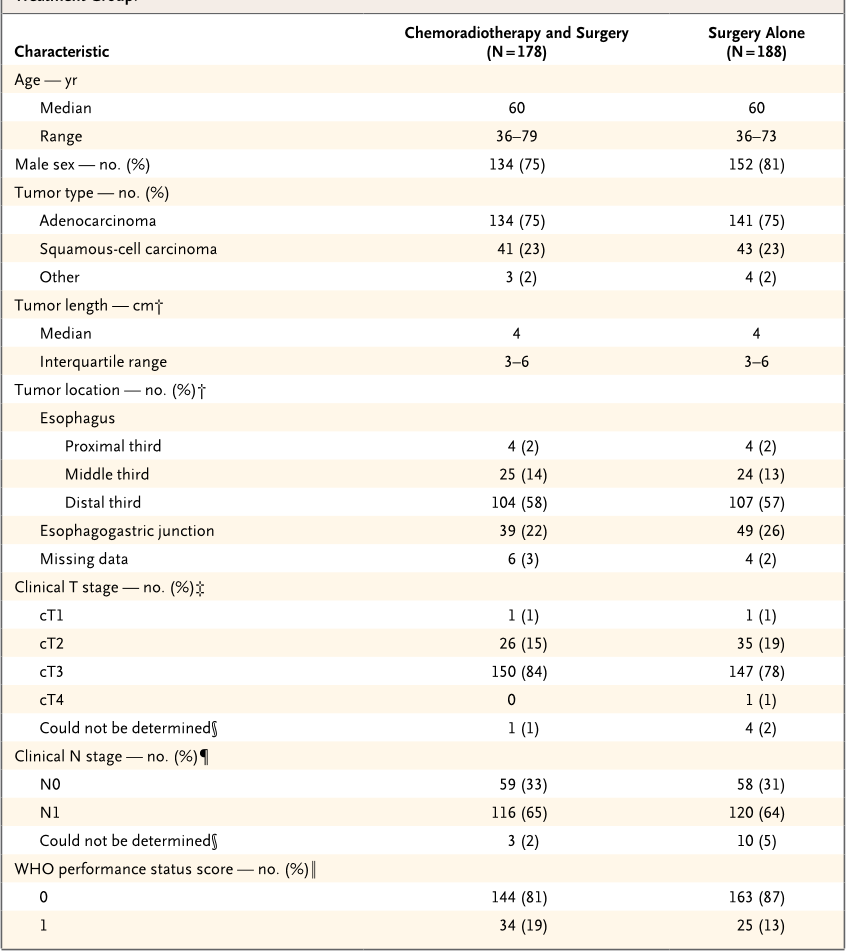
Table 1. Characteristics of Patients with Resectable Esophageal or Esophagogastric-Junction Cancer, According to
Treatment Group.*
Characteristic
Chemoradiotherapy and Surgery
(N = 178)
Surgery Alone
(N = 188)
Age — yr
Median 60 60
Range 36–79 36–73
Male sex — no. (%) 134 (75) 152 (81)
Tumor type — no. (%)
Adenocarcinoma 134 (75) 141 (75)
Squamous-cell carcinoma 41 (23) 43 (23)
Other 3 (2) 4 (2)
Tumor length — cm†
Median 4 4
Interquartile range 3–6 3–6
Tumor location — no. (%)†
Esophagus
Proximal third 4 (2) 4 (2)
Middle third 25 (14) 24 (13)
Distal third 104 (58) 107 (57)
Esophagogastric junction 39 (22) 49 (26)
Missing data 6 (3) 4 (2)
Clinical T stage — no. (%)‡
cT1 1 (1) 1 (1)
cT2 26 (15) 35 (19)
cT3 150 (84) 147 (78)
cT4 0 1 (1)
Could not be determined§ 1 (1) 4 (2)
Clinical N stage — no. (%)¶
N0 59 (33) 58 (31)
N1 116 (65) 120 (64)
Could not be determined§ 3 (2) 10 (5)
WHO performance status score — no. (%)‖
0 144 (81) 163 (87)
1 34 (19) 25 (13)
* Percentages may not add up to 100 because of rounding. WHO denotes World Health Organization.
Tumor length and location were determined by means of endoscopy.
Clinical tumor (cT) stage was assessed by means of endoscopic ultrasonography or computed tomography (CT) and was
classified according to the International Union against Cancer (UICC) tumor–node–metastasis (TNM) classification.
9
§ This category included patients in whom the tumor could not be fully investigated by means of a transducer for endoscopic
ultrasonography owing to a stenosis caused by the tumor.
Clinical lymph-node (N) stage was assessed by means of endoscopic ultrasonography, CT, or
18
F-fluorodeoxyglucose
positron-emission tomography and was classified according to UICC TNM classification.
9
WHO performance status scores are on a scale of 0 to 5, with lower numbers indicating better performance status; 0 indi-
cates fully active, and 1 unable to carry out heavy physical work.
The New England Journal of Medicine
Downloaded from nejm.org at VRIJE UNIVERSITEIT on October 29, 2013. For personal use only. No other uses without permission.
Copyright © 2012 Massachusetts Medical Society. All rights reserved.

The
ne w engl and jour nal
o f
medicine
n engl j med 366;22 nejm.org may 31, 2012
2078
the date of randomization until death. All data
collected through December 2010 were included
in the analysis, which guaranteed a potential
minimal follow-up of 2 years.
The Kaplan–Meier method was used to esti-
mate survival, with the log-rank test to deter-
mine significance. A Cox proportional-hazards
model was used to estimate the treatment effect
with adjustment for prognostic factors for sur-
vival. Moreover, Cox models were used to iden-
tify possible interactions in treatment effect
between subgroups, both with and without
adjustment for prognostic factors. Subgroups
were predefined according to sex, histologic
subtype of tumor, clinical N stage, and WHO
performance score. Statistical analysis was per-
formed with the use of SPSS software, version
17.0 (SPSS).
RESULTS
CHARACTERISTICS OF THE PATIENTS
From March 2004 through December 2008, we
enrolled 368 patients in the study, of whom 180
were randomly assigned to the chemoradiother-
apy–surgery group, and 188 to the surgery group.
Two patients who were randomly assigned to
the chemoradiotherapy–surgery group withdrew
consent and were not included in the analysis
(Fig. 1).
Prognostic factors were well balanced be-
tween the two treatment groups (
Table 1
). In
both groups, the median age was 60 years; 134
of 178 patients (75%) in the chemoradiotherapy–
surgery group were men, as compared with 152
of 188 patients (81%) in the surgery group. Most
patients (275 of 366 [75%]) had an adenocarci-
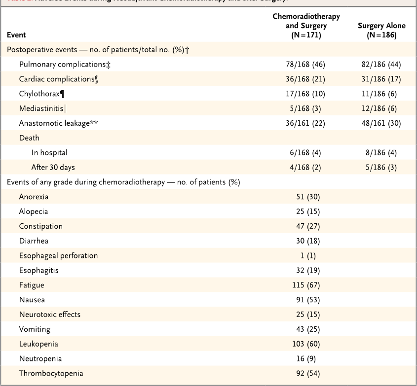
Table 2. Adverse Events during Neoadjuvant Chemoradiotherapy and after Surgery.*
Event
Chemoradiotherapy
and Surgery
(N = 171)
Surgery Alone
(N = 186)
Postoperative events — no. of patients/total no. (%)†
Pulmonary complications‡ 78/168 (46) 82/186 (44)
Cardiac complications§ 36/168 (21) 31/186 (17)
Chylothorax¶ 17/168 (10) 11/186 (6)
Mediastinitis‖ 5/168 (3) 12/186 (6)
Anastomotic leakage** 36/161 (22) 48/161 (30)
Death
In hospital 6/168 (4) 8/186 (4)
After 30 days 4/168 (2) 5/186 (3)
Events of any grade during chemoradiotherapy — no. of patients (%)
Anorexia 51 (30)
Alopecia 25 (15)
Constipation 47 (27)
Diarrhea 30 (18)
Esophageal perforation 1 (1)
Esophagitis 32 (19)
Fatigue 115 (67)
Nausea 91 (53)
Neurotoxic effects 25 (15)
Vomiting 43 (25)
Leukopenia 103 (60)
Neutropenia 16 (9)
Thrombocytopenia 92 (54)
The New England Journal of Medicine
Downloaded from nejm.org at VRIJE UNIVERSITEIT on October 29, 2013. For personal use only. No other uses without permission.
Copyright © 2012 Massachusetts Medical Society. All rights reserved.
Citations
More filters
Journal ArticleDOI
TL;DR: Long-term follow-up confirms the overall survival benefits for neoadjuvant chemoradiotherapy in patients with clinically resectable, locally advanced cancer of the oesophagus or Oesophagogastric junction and shows a significant increase in 5-year overall survival.
Abstract: Summary Background Initial results of the ChemoRadiotherapy for Oesophageal cancer followed by Surgery Study (CROSS) comparing neoadjuvant chemoradiotherapy plus surgery versus surgery alone in patients with squamous cell carcinoma and adenocarcinoma of the oesophagus or oesophagogastric junction showed a significant increase in 5-year overall survival in favour of the neoadjuvant chemoradiotherapy plus surgery group after a median of 45 months' follow-up. In this Article, we report the long-term results after a minimum follow-up of 5 years. Methods Patients with clinically resectable, locally advanced cancer of the oesophagus or oesophagogastric junction (clinical stage T1N1M0 or T2–3N0–1M0, according to the TNM cancer staging system, sixth edition) were randomly assigned in a 1:1 ratio with permuted blocks of four or six to receive either weekly administration of five cycles of neoadjuvant chemoradiotherapy (intravenous carboplatin [AUC 2 mg/mL per min] and intravenous paclitaxel [50 mg/m 2 of body-surface area] for 23 days) with concurrent radiotherapy (41·4 Gy, given in 23 fractions of 1·8 Gy on 5 days per week) followed by surgery, or surgery alone. The primary endpoint was overall survival, analysed by intention-to-treat. No adverse event data were collected beyond those noted in the initial report of the trial. This trial is registered with the Netherlands Trial Register, number NTR487, and has been completed. Findings Between March 30, 2004, and Dec 2, 2008, 368 patients from eight participating centres (five academic centres and three large non-academic teaching hospitals) in the Netherlands were enrolled into this study and randomly assigned to the two treatment groups: 180 to surgery plus neoadjuvant chemoradiotherapy and 188 to surgery alone. Two patients in the neoadjuvant chemoradiotherapy group withdrew consent, so a total of 366 patients were analysed (178 in the neoadjuvant chemoradiotherapy plus surgery group and 188 in the surgery alone group). Of 171 patients who received any neoadjuvant chemoradiotherapy in this group, 162 (95%) were able to complete the entire neoadjuvant chemoradiotherapy regimen. After a median follow-up for surviving patients of 84·1 months (range 61·1–116·8, IQR 70·7–96·6), median overall survival was 48·6 months (95% CI 32·1–65·1) in the neoadjuvant chemoradiotherapy plus surgery group and 24·0 months (14·2–33·7) in the surgery alone group (HR 0·68 [95% CI 0·53–0·88]; log-rank p=0·003). Median overall survival for patients with squamous cell carcinomas was 81·6 months (95% CI 47·2–116·0) in the neoadjuvant chemoradiotherapy plus surgery group and 21·1 months (15·4–26·7) in the surgery alone group (HR 0·48 [95% CI 0·28–0·83]; log-rank p=0·008); for patients with adenocarcinomas, it was 43·2 months (24·9–61·4) in the neoadjuvant chemoradiotherapy plus surgery group and 27·1 months (13·0–41·2) in the surgery alone group (HR 0·73 [95% CI 0·55–0·98]; log-rank p=0·038). Interpretation Long-term follow-up confirms the overall survival benefits for neoadjuvant chemoradiotherapy when added to surgery in patients with resectable oesophageal or oesophagogastric junctional cancer. This improvement is clinically relevant for both squamous cell carcinoma and adenocarcinoma subtypes. Therefore, neoadjuvant chemoradiotherapy according to the CROSS trial followed by surgical resection should be regarded as a standard of care for patients with resectable locally advanced oesophageal or oesophagogastric junctional cancer. Funding Dutch Cancer Foundation (KWF Kankerbestrijding).

1,703 citations

Journal ArticleDOI
TL;DR: In this article, the safety and efficacy of the docetaxel-based triplet FLOT (fluorouracil plus leucovorin, oxaliplatin, and doceteaxel) as a perioperative therapy for patients with locally advanced, resectable tumours was reported.

1,218 citations

Journal ArticleDOI
TL;DR: It is reported that circulating tumor cells become trapped within NETs in vitro under static and dynamic conditions and NETs are identified as potential therapeutic targets in the context of systemic infection.
Abstract: The majority of patients with cancer undergo at least one surgical procedure as part of their treatment. Severe postsurgical infection is associated with adverse oncologic outcomes; however, the mechanisms underlying this phenomenon are unclear. Emerging evidence suggests that neutrophils, which function as the first line of defense during infections, facilitate cancer progression. Neutrophil extracellular traps (NETs) are extracellular neutrophil-derived DNA webs released in response to inflammatory cues that trap and kill invading pathogens. The role of NETs in cancer progression is entirely unknown. We report that circulating tumor cells become trapped within NETs in vitro under static and dynamic conditions. In a murine model of infection using cecal ligation and puncture, we demonstrated microvascular NET deposition and consequent trapping of circulating lung carcinoma cells within DNA webs. NET trapping was associated with increased formation of hepatic micrometastases at 48 hours and gross metastatic disease burden at 2 weeks following tumor cell injection. These effects were abrogated by NET inhibition with DNAse or a neutrophil elastase inhibitor. These findings implicate NETs in the process of cancer metastasis in the context of systemic infection and identify NETs as potential therapeutic targets.

921 citations


Cites background from "Preoperative Chemoradiotherapy for ..."

  • ...Currently, locoregional control in the form of complete oncologic resection remains an essential curative modality for nearly all solid tumors and provides improved overall and disease-free survival (2, 4, 5)....

    [...]

Journal ArticleDOI
TL;DR: In the EU, the highest age-standardised incidence rates for oesophageal cancer are in the Netherlands for men and the UK for women, and variation between countries is high and may reflect different prevalence of risk factors, use of screening and diagnostic methods.

810 citations

Journal ArticleDOI
TL;DR: This selection from the NCCN Guidelines for Esophageal and Esophagogastric Junction Cancers focuses on recommendations for the management of locally advanced and metastatic adenocarcinoma of the esophagus and EGJ.
Abstract: Esophageal cancer is the sixth leading cause of cancer-related deaths worldwide. Squamous cell carcinoma is the most common histology in Eastern Europe and Asia, and adenocarcinoma is most common in North America and Western Europe. Surgery is a major component of treatment of locally advanced resectable esophageal and esophagogastric junction (EGJ) cancer, and randomized trials have shown that the addition of preoperative chemoradiation or perioperative chemotherapy to surgery significantly improves survival. Targeted therapies including trastuzumab, ramucirumab, and pembrolizumab have produced encouraging results in the treatment of patients with advanced or metastatic disease. Multidisciplinary team management is essential for all patients with esophageal and EGJ cancers. This selection from the NCCN Guidelines for Esophageal and Esophagogastric Junction Cancers focuses on recommendations for the management of locally advanced and metastatic adenocarcinoma of the esophagus and EGJ.

710 citations

References
More filters
Journal ArticleDOI
TL;DR: Although the study was closed early and statistical significance was not achieved, results point to a survival advantage for preoperative chemoradiotherapy compared with preoperative chemotherapy in adenocarcinomas of the esophagogastric junction.
Abstract: Purpose Preoperative chemotherapy is an accepted standard in the treatment of localized esophagogastric adenocarcinoma. Adding radiation therapy to preoperative chemotherapy appears promising, but its definitive value remains unknown. Patients and Methods Patients with locally advanced (uT3-4NXM0) adenocarcinoma of the lower esophagus or gastric cardia were randomly allocated to one of two treatment groups: induction chemotherapy (15 weeks) followed by surgery (arm A); or chemotherapy (12 weeks) followed by chemoradiotherapy (3 weeks) followed by surgery (arm B). Primary outcome was overall survival time. A total of 354 patients were needed to detect a 10% increase in 3-year survival from 25% to 35% by addition of radiation therapy. The study was prematurely closed due to low accrual. Results The median observation time was 46 months. A total of 126 patients were randomly assigned and 119 eligible patients were evaluated. The number of patients undergoing complete tumor resection was not different between treatment groups (69.5% v 71.5%). Patients in arm B had a significant higher probability of showing pathologic complete response (15.6% v 2.0%) or tumor-free lymph nodes (64.4% v 37.7%) at resection. Preoperative radiation therapy improved 3-year survival rate from 27.7% to 47.4% (log-rank P .07, hazard ratio adjusted for randomization strata variables 0.67, 95% CI, 0.41 to 1.07). Postoperative mortality was nonsignificantly increased in the chemoradiotherapy group (10.2% v 3.8%; P .26). Conclusion Although the study was closed early and statistical significance was not achieved, results point to a survival advantage for preoperative chemoradiotherapy compared with preoperative chemotherapy in adenocarcinomas of the esophagogastric junction.

855 citations

Journal ArticleDOI
01 Jul 2004-Gut
TL;DR: In patients with resectable oesophageal cancer, chemoradiotherapy plus surgery significantly reduces three year mortality compared with surgery alone, however, postoperative mortality was significantly increased by neoadjuvant cheMoradiotherapy.
Abstract: Background: The benefit of neoadjuvant chemoradiotherapy in oesophageal cancer has been extensively studied but data on survival are still equivocal. Objective: To assess the effectiveness of chemoradiotherapy followed by surgery in the reduction of mortality in patients with resectable oesophageal cancer. Methods: Computerised bibliographic searches of MEDLINE and CANCERLIT (1970–2002) were supplemented with hand searches of reference lists. Study selection: Studies were included if they were randomised controlled trials (RCTs) comparing preoperative chemoradiotherapy plus surgery with surgery alone, and if they included patients with resectable histologically proven oesophageal cancer without metastatic disease. Six eligible RCTs were identified and included in the meta-analysis. Data extraction: Data on study populations, interventions, and outcomes were extracted from each RCT according to the intention to treat method by three independent observers and combined using the DerSimonian and Laird method. Results: Chemoradiotherapy plus surgery compared with surgery alone significantly reduced the three year mortality rate (odds ratio (OR) 0.53 (95% confidence interval (CI) 0.31–0.93); p = 0.03) (number needed to treat = 10). Pathological examination showed that preoperative chemoradiotherapy downstaged the tumour (that is, less advanced stage at pathological examination at the time of surgery) compared with surgery alone (OR 0.43 (95% CI 0.26–0.72); p = 0.001). The risk for postoperative mortality was higher in the chemoradiotherapy plus surgery group (OR 2.10 (95% CI 1.18–3.73); p = 0.01). Conclusions: In patients with resectable oesophageal cancer, chemoradiotherapy plus surgery significantly reduces three year mortality compared with surgery alone. However, postoperative mortality was significantly increased by neoadjuvant chemoradiotherapy. Further large scale multicentre RCTs may prove useful to substantiate the benefit on overall survival.

546 citations

Journal ArticleDOI
TL;DR: In patients with squamous-cell esophageal cancer, preoperative chemoradiotherapy did not improve overall survival, but it did prolong disease-free survival and survival free of local disease.
Abstract: BACKGROUND We conducted a multicenter, randomized trial to compare preoperative chemoradiotherapy followed by surgery with surgery alone in patients with stage I and II squamous-cell cancer of the esophagus. METHODS The preoperative combined therapy consisted of two one-week courses; each involved radiotherapy, in a dose of 18.5 Gy delivered in five fractions of 3.7 Gy each, and 80 mg of cisplatin per square meter of body-surface area, administered 0 to 2 days before the first day of radiotherapy. The surgical plan included one-stage en bloc esophagectomy and proximal gastrectomy by the abdominal and right thoracic routes, to be performed immediately after randomization in the group assigned to surgery alone and two to four weeks after the completion of preoperative chemoradiotherapy in the group assigned to combined therapy. RESULTS A total of 297 patients entered the study; 11 were found to be ineligible, and 4 were lost to follow-up. Of the remaining 282, 139 were assigned to surgery alone and 143 to combined therapy. After a median follow-up of 55.2 months, no significant difference in overall survival was observed; the median survival was 18.6 months for both groups. As compared with the group treated with surgery alone, the group treated preoperatively had longer disease-free survival (P=0.003), a longer interval free of local disease (P=0.01), a lower rate of cancer-related deaths (P=0.002), and a higher frequency of curative resection (P=0.017). However, there were more postoperative deaths (P=0.012) in the group treated preoperatively with chemoradiotherapy. Three prognostic factors were found to influence survival in a multivariate analysis: the disease stage, based on computed tomography; the location of the tumor; and whether the surgical resection was curative. CONCLUSIONS In patients with squamous-cell esophageal cancer, preoperative chemoradiotherapy did not improve overall survival, but it did prolong disease-free survival and survival free of local disease.

545 citations

Journal ArticleDOI
01 Apr 2005-Cancer
TL;DR: In patients with locoregional carcinoma of the esophagus or esophagogastric junction who underwent preoperative chemoradiation, it is unclear whether survival was better predicted by pretherapy clinical stage or by posttherapy pathologic stage.
Abstract: BACKGROUND In patients with locoregional carcinoma of the esophagus or esophagogastric junction who underwent preoperative chemoradiation, it is unclear whether survival was better predicted by pretherapy clinical stage or by posttherapy pathologic stage METHODS The authors studied 235 consecutive patients with pretherapy clinical Stage II, III, or IVA (according to American Joint Committee on Cancer criteria) carcinoma of the esophagus or esophagogastric junction who were treated with chemoradiation followed by esophagectomy Posttherapy cancer status was classified using pathologic stage and semiquantitative assessment of residual carcinoma Clinicopathologic features, residual carcinoma status, and pretherapy and posttherapy stage were compared with disease-free and overall survival RESULTS Posttherapy pathologic stage was Stage 0 in 29% of patients, Stage I in 11% of patients, Stage II in 34% of patients, Stage III in 20% of patients, and Stage IV in 6% of patients Cancer downstaging occurred in 56% of patients In univariate analysis, disease-free and overall survival were predicted by posttherapy pathologic stage (both with P < 0001), margin status (P = 0002 and P = 001, respectively), extent of residual carcinoma (both with P < 0001), and downstaging (both with P = 0001), but not by age, gender, type of cancer, pretherapy clinical stage, or preoperative regimen However, in multivariate analysis, disease-free and overall survival were independently predicted by posttherapy pathologic stage (both with P = 002) Extent of residual carcinoma was a marginally significant predictor of overall survival (P = 004) CONCLUSIONS Posttherapy pathologic stage was the best available predictor of outcome for patients with locoregional carcinoma of the esophagus or esophagogastric junction who underwent chemoradiation therapy followed by esophagectomy The findings in the current study supported the concept of downstaging by preoperative therapy Cancer 2005 © 2005 American Cancer Society

426 citations

Related Papers (5)