scispace - formally typeset
Search or ask a question
Posted ContentDOI

Selective cleavage of ncRNA and antiviral activity by human RNase2/EDN in a macrophage infection model

31 Aug 2021-bioRxiv (Cold Spring Harbor Laboratory)-
TL;DR: In this article, the authors investigated the role of RNase2 in antiviral host defence against RSV infection and showed that the cleavage sites at the anticodon loops and secondarily at D-loops are the most prominent sites of cleavage.
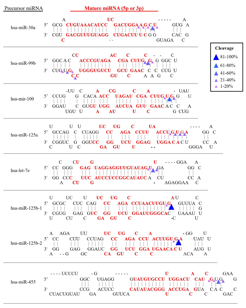
Abstract: RNase2, also named the Eosinophil derived Neurotoxin (EDN), is one of the main proteins secreted by the eosinophil secondary granules. RNase2 is also expressed in other leukocyte cells and is the member of the human ribonuclease A family most abundant in macrophages. The protein is endowed with a high ribonucleolytic activity and participates in the host antiviral activity. Although RNase2 displays a broad antiviral activity, it is mostly associated to the targeting of single stranded RNA viruses. To explore RNase2 mechanism of action in antiviral host defence we knocked out RNase2 expression in the THP1 monocyte cell line and characterized the cell response to human Respiratory Syncytial Virus (RSV). We observed that RSV infection induced the RNase2 expression and protein secretion in THP1 macrophage-derived cells, whereas the knockout (KO) of RNase2 resulted in higher RSV burden and reduced cell viability. Next, by means of the cP-RNAseq methodology, which uniquely amplifies the RNA 2939cyclic-phosphate-end products released by an endonuclease cleavage, we compared the ncRNA population in native and RNase2-KO cell lines. Among the ncRNAs accumulated in WT versus KO cells, we found mostly tRNA-derived fragments and secondly miRNAs. Analysis of the differential sequence coverage of tRNAs molecules in native and KO cells identified fragments derived from only few parental tRNAs, revealing a predominant cleavage at anticodon loops and secondarily at D-loops. Inspection of cleavage region identified U/C and A, at 59 and 39 sides of cleavage sites respectively (namely RNase B1 and B2 base binding subsites). Likewise, only few selected miRNAs were significantly more abundant in WT versus RNase2-KO cells, with cleavage sites located at the end of stem regions with predominance for pyrimidines at B1 but following an overall less defined nucleotide specificity. Complementarily, by screening of a tRF/tiRNA PCR array we identified an enriched population of tRNA-derived fragments associated to RNase2 expression and RSV infection. The present results confirm the contribution of the protein in macrophage response against virus infection and provide the first evidence of its cleavage selectivity against ncRNA population. A better understanding of the mechanism of action of RNase2 recognition of cellular RNA during the antiviral host defence should pave the basis for the design of novel antiviral drugs.

Summary (1 min read)

Jump to: [Introduction][Results][Discussion] and [Conclusions]

Introduction

  • Human RNase2 is a secretory protein expressed in leukocytes with a reported antiviral activity against single stranded RNA viruses [1,2].
  • Recently, presence of eosinophils and their associated RNases has been correlated to the prognosis of COVID patients [15–17].
  • RNase2 was proposed to have a role in the host response against the single stranded RNA virus [22] and early studies observed that RNase2 can directly target the RSV virion [12].

Results

  • RSV infection activated the expression of RNase2 in macrophages RSV virus stock was obtained at a titration of 2.8×10⁶ TCID50/mL, as previously described [25] and THP1 macrophages were exposed to the RSV at a selected MOI of 1:1 up to 72h post of infection (poi).
  • Moreover, the total absence of secreted RNase2 by THP1 cells was confirmed by ELISA assay in culture supernatant for KO28 line (Fig 2C).
  • At 24h, RNase2 KO macrophages had significantly more intracellular RSV than WT macrophages.
  • Following RNAseq amplification, the sequence libraries were inspected by differential enrichment analysis.
  • Therefore, their data is the first report of the specific release of tRNA fragments associated to RNase2 expression.

Discussion

  • Expression of human RNase2 is widely distributed in diverse body tissues such as liver and spleen together with leukocyte cells [2].
  • In contrast, using the same infection model and experimental protocol, the authors observe here how RSV infection significantly activate both the expression and protein secretion of RNase2 in THP1 macrophage derived cells (Fig 1).
  • In addition, the cleavage of tRNAs by RNases would probably be modulated by the presence of regulatory proteins within the cell [60].
  • This might explain some of the differences observed in the identified fragments when comparing the screening of the tRFs array and the amplified sequences by the Cp-RNAseq methodology, which only amplifies the products of an endonuclease cleavage.
  • Their results confirm that RNase2 targets ncRNA and releases specific miRNAs and tRFs.

Conclusions

  • This is the first report of RNase2 selective targeting of ncRNA.
  • Comparison of native and knockout THP1-derived macrophages in a RSV infection model confirms the RNase involvement in the cell host antiviral defence.
  • By amplification of 2’3Cp end RNA products, the authors have identified the tRNA fragments and miRNAs associated to RNase2 cleavage.
  • The analysis of RNA recognition regions reveals the RNA base composition at the 5’ and 3’ of cleavage site.
  • Further work is mandatory for an unambiguous pattern assignment towards the understanding of how RNase2 can shape the ncRNA population and its role to fight viral infection.

Did you find this useful? Give us your feedback

Figures (8)

Content maybe subject to copyright    Report

1
Selective cleavage of ncRNA and antiviral activity by human
RNase2/EDN in a macrophage infection model
Lu Lu
1,2#*
, Jiarui Li
1*
, Ranlei Wei
3
, Irene Guidi
1
, Luca Cozzuto
4
, Julia
Ponomarenko
4
, Guillem Prats-Ejarque
1
and Ester Boix
1#
1
Department of Biochemistry and Molecular Biology, Faculty of Biosciences, Universitat
Autonomous de Barcelona, Cerdanyola del Vallès, Spain
2
College of Animal Science and Technology, Sichuan Agricultural University, Chengdu,
Sichuan, China
3
Center of Precision Medicine& Precision Medicine Key Laboratory of Sichuan Province,
West China Hospital, Sichuan University, Chengdu, China
4
Bioinformatic Unit. Centre de Regulació Genòmica (CRG), Barcelona, Spain
*Both authors contributed equally to this work
#
Corresponding authors: Ester Boix, Ester.Boix@uab.es ; Lu Lu, lu.lu@sicau.edu.cn
(which was not certified by peer review) is the author/funder. All rights reserved. No reuse allowed without permission.
The copyright holder for this preprintthis version posted August 31, 2021. ; https://doi.org/10.1101/2021.08.30.458223doi: bioRxiv preprint

2
Abstract
RNase2, also named the Eosinophil derived Neurotoxin (EDN), is one of the
main proteins secreted by the eosinophil secondary granules. RNase2 is also expressed in
other leukocyte cells and is the member of the human ribonuclease A family most
abundant in macrophages. The protein is endowed with a high ribonucleolytic activity
and participates in the host antiviral activity. Although RNase2 displays a broad antiviral
activity, it is mostly associated to the targeting of single stranded RNA viruses. To explore
RNase2 mechanism of action in antiviral host defence we knocked out RNase2 expression
in the THP1 monocyte cell line and characterized the cell response to human Respiratory
Syncytial Virus (RSV). We observed that RSV infection induced the RNase2 expression
and protein secretion in THP1 macrophage-derived cells, whereas the knockout (KO) of
RNase2 resulted in higher RSV burden and reduced cell viability. Next, by means of the
cP-RNAseq methodology, which uniquely amplifies the RNA 2’3’cyclic-phosphate-end
products released by an endonuclease cleavage, we compared the ncRNA population in
native and RNase2-KO cell lines. Among the ncRNAs accumulated in WT versus KO
cells, we found mostly tRNA-derived fragments and secondly miRNAs. Analysis of the
differential sequence coverage of tRNAs molecules in native and KO cells identified
fragments derived from only few parental tRNAs, revealing a predominant cleavage at
anticodon loops and secondarily at D-loops. Inspection of cleavage region identified U/C
and A, at 5’ and 3’ sides of cleavage sites respectively (namely RNase B1 and B2 base
binding subsites). Likewise, only few selected miRNAs were significantly more abundant
in WT versus RNase2-KO cells, with cleavage sites located at the end of stem regions
with predominance for pyrimidines at B1 but following an overall less defined nucleotide
specificity.
(which was not certified by peer review) is the author/funder. All rights reserved. No reuse allowed without permission.
The copyright holder for this preprintthis version posted August 31, 2021. ; https://doi.org/10.1101/2021.08.30.458223doi: bioRxiv preprint

3
Complementarily, by screening of a tRF&tiRNA PCR array we identified an
enriched population of tRNA-derived fragments associated to RNase2 expression and
RSV infection. The present results confirm the contribution of the protein in macrophage
response against virus infection and provide the first evidence of its cleavage selectivity
against ncRNA population. A better understanding of the mechanism of action of RNase2
recognition of cellular RNA during the antiviral host defence should pave the basis for
the design of novel antiviral drugs.
Short title: RNase2-induced specific ncRNA cleavage
Keywords: RNase2, EDN, macrophage, antiviral, RSV, ncRNA, tRNAs, tRFs
(which was not certified by peer review) is the author/funder. All rights reserved. No reuse allowed without permission.
The copyright holder for this preprintthis version posted August 31, 2021. ; https://doi.org/10.1101/2021.08.30.458223doi: bioRxiv preprint

4
Introduction
Human RNase2 is a secretory protein expressed in leukocytes with a reported
antiviral activity against single stranded RNA viruses [1,2]. RNase2 is one of the main
components of the eosinophil secondary granule matrix. The protein, upon its discovery,
was named the Eosinophil Derived Neurotoxin (EDN), due to its ability to induce the
Gordon phenomenon when injected into Guinea pigs [3–8].Apart from eosinophils,
RNase2 is also expressed in other leukocyte cell types, such as neutrophils and
monocytes, together with epithelial cells, liver and spleen [8–10]. The protein belongs to
the ribonuclease A superfamily, a family of secretory RNases that participate in the host
response and combine a direct action against a wide range of pathogens with diverse
immunomodulation properties [2,11].
RNase2 stands out for its high catalytic activity against single stranded RNA and
its efficiency against several viral types, such as rhinoviruses, adenoviruses and
retroviruses, including HIV [12–14]. Recently, presence of eosinophils and their
associated RNases has been correlated to the prognosis of COVID patients [15–17]. On
the contrary, no action is reported against the tested bacterial species [12,18,19]. In
particular, among respiratory viruses, which activate eosinophil recruitment and
degranulation, the human Respiratory Syncytial Virus (RSV), which is the principal cause
of death in infants [20], is probably the most studied model for RNase2 antiviral action.
Indeed, RNase2 levels have predictive value for the development of recurrent wheezing
post-RSV bronchiolitis [21]. RNase2 was proposed to have a role in the host response
against the single stranded RNA virus [22] and early studies observed that RNase2 can
directly target the RSV virion [12]. Interestingly, the protein ribonucleolytic activity is
required to remove the RSV genome, but some structural specificity for RNase2 is
(which was not certified by peer review) is the author/funder. All rights reserved. No reuse allowed without permission.
The copyright holder for this preprintthis version posted August 31, 2021. ; https://doi.org/10.1101/2021.08.30.458223doi: bioRxiv preprint

5
mandatory, as other family homologues endowed with a higher catalytic activity are
devoid of antiviral activity [23].
In our previous work using a macrophage infection model, we observed that
RNase2 is the most abundantly expressed RNaseA superfamily member in the monocytic
THP1 cell line [24]. To broaden the knowledge of the immunomodulatory role and
potential targeting of cellular RNA population by RNase2 in human macrophages, we
built an RNase2-knockout THP1 monocyte cell line using CRIPSR/Cas9 (clustered
regularly interspaced short palindromic repeats) gene editing tool. Transcriptome of the
RNase2 knockout with the unedited THP1-derived macrophage cells revealed that the top
differently expressed pathways are associated to antiviral host defence (Lu et al., in
preparation). Here, we explored the protein antiviral action by characterization of both
THP1 native and RNase2-KO cell lines infected with RSV. The comparative infection
study indicated that the knockout of RNase2 in THP1- derived macrophages resulted in
a heavier RSV titre and reduced cell survival. Next, we analysed the total non-coding
RNA (ncRNA) population by amplification of 2’3’-cyclic phosphate ends using the cp-
RNAseq methodology and by screening a library array of tRNA-derived fragments.
Results proved that RNase2 expression in macrophage correlates to a selective ncRNA
cleavage pattern.
(which was not certified by peer review) is the author/funder. All rights reserved. No reuse allowed without permission.
The copyright holder for this preprintthis version posted August 31, 2021. ; https://doi.org/10.1101/2021.08.30.458223doi: bioRxiv preprint

References
More filters
Journal ArticleDOI
TL;DR: This work presents DESeq2, a method for differential analysis of count data, using shrinkage estimation for dispersions and fold changes to improve stability and interpretability of estimates, which enables a more quantitative analysis focused on the strength rather than the mere presence of differential expression.
Abstract: In comparative high-throughput sequencing assays, a fundamental task is the analysis of count data, such as read counts per gene in RNA-seq, for evidence of systematic changes across experimental conditions. Small replicate numbers, discreteness, large dynamic range and the presence of outliers require a suitable statistical approach. We present DESeq2, a method for differential analysis of count data, using shrinkage estimation for dispersions and fold changes to improve stability and interpretability of estimates. This enables a more quantitative analysis focused on the strength rather than the mere presence of differential expression. The DESeq2 package is available at http://www.bioconductor.org/packages/release/bioc/html/DESeq2.html .

47,038 citations

Journal ArticleDOI
TL;DR: Bowtie extends previous Burrows-Wheeler techniques with a novel quality-aware backtracking algorithm that permits mismatches and can be used simultaneously to achieve even greater alignment speeds.
Abstract: Bowtie is an ultrafast, memory-efficient alignment program for aligning short DNA sequence reads to large genomes. For the human genome, Burrows-Wheeler indexing allows Bowtie to align more than 25 million reads per CPU hour with a memory footprint of approximately 1.3 gigabytes. Bowtie extends previous Burrows-Wheeler techniques with a novel quality-aware backtracking algorithm that permits mismatches. Multiple processor cores can be used simultaneously to achieve even greater alignment speeds. Bowtie is open source http://bowtie.cbcb.umd.edu.

20,335 citations

Journal ArticleDOI
TL;DR: This work presents HTSeq, a Python library to facilitate the rapid development of custom scripts for high-throughput sequencing data analysis, and presents htseq-count, a tool developed with HTSequ that preprocesses RNA-Seq data for differential expression analysis by counting the overlap of reads with genes.
Abstract: Motivation: A large choice of tools exists for many standard tasks in the analysis of high-throughput sequencing (HTS) data. However, once a project deviates from standard workflows, custom scripts are needed. Results: We present HTSeq, a Python library to facilitate the rapid development of such scripts. HTSeq offers parsers for many common data formats in HTS projects, as well as classes to represent data, such as genomic coordinates, sequences, sequencing reads, alignments, gene model information and variant calls, and provides data structures that allow for querying via genomic coordinates. We also present htseq-count, a tool developed with HTSeq that preprocesses RNA-Seq data for differential expression analysis by counting the overlap of reads with genes. Availability and implementation: HTSeq is released as an opensource software under the GNU General Public Licence and available from http://www-huber.embl.de/HTSeq or from the Python Package Index at https://pypi.python.org/pypi/HTSeq. Contact: sanders@fs.tum.de

15,744 citations

Journal ArticleDOI
TL;DR: The miRBase database aims to provide integrated interfaces to comprehensive microRNA sequence data, annotation and predicted gene targets, and acts as an independent arbiter of microRNA gene nomenclature.
Abstract: The miRBase database aims to provide integrated interfaces to comprehensive microRNA sequence data, annotation and predicted gene targets. miRBase takes over functionality from the microRNA Registry and fulfils three main roles: the miRBase Registry acts as an independent arbiter of microRNA gene nomenclature, assigning names prior to publication of novel miRNA sequences. miRBase Sequences is the primary online repository for miRNA sequence data and annotation. miRBase Targets is a comprehensive new database of predicted miRNA target genes. miRBase is available at http://microrna.sanger.ac.uk/.

4,629 citations

Journal ArticleDOI
TL;DR: Since the discovery in 1993 of the first small silencing RNA, a dizzying number of small RNA classes have been identified, including microRNAs (miRNAs), small interfering RNAs (siRNAs) and Piwi-interacting RNAs.
Abstract: Since the discovery in 1993 of the first small silencing RNA, a dizzying number of small RNA classes have been identified, including microRNAs (miRNAs), small interfering RNAs (siRNAs) and Piwi-interacting RNAs (piRNAs). These classes differ in their biogenesis, their modes of target regulation and in the biological pathways they regulate. There is a growing realization that, despite their differences, these distinct small RNA pathways are interconnected, and that small RNA pathways compete and collaborate as they regulate genes and protect the genome from external and internal threats.

2,266 citations

Frequently Asked Questions (15)
Q1. What are the contributions mentioned in the paper "Selective cleavage of ncrna and antiviral activity by human rnase2/edn in a macrophage infection model" ?

In this paper, the authors showed that RNase2 expression in macrophages correlates with a selective ncRNA cleavage pattern. 

Further work is mandatory for an unambiguous pattern assignment towards the understanding of how RNase2 can shape the ncRNA population and its role to fight viral infection. 

Among the blood cell types, RNase2 is particularly abundant in monocytes [11], which are key contributors to host defence against pathogens. 

RNase2 was proposed to have a role in the host response against the single stranded RNA virus [22] and early studies observed that RNase2 can directly target the RSV virion [12]. 

Accumulation of miRNAs can be toxic to the cells, due to their potential interference with the translation of essential proteins. 

Particular interest should be drawn to the new identified tRNA fragments associated to RNase2 and absent from the commercial library array, which represent potential new regulatory elements for future studies. 

Increasing data demonstrates that small noncoding RNAs (ncRNAs) play important roles in regulating antiviral innate immune responses [34–36]. 

RSV infection activated the expression of RNase2 inmacrophagesRSV virus stock was obtained at a titration of 2.8×10⁶ TCID50/mL, as previouslydescribed [25] and THP1 macrophages were exposed to the RSV at a selected MOI of 1:1 up to 72h post of infection (poi). 

In their previous work using a macrophage infection model, the authors observed thatRNase2 is the most abundantly expressed RNaseA superfamily member in the monocytic THP1 cell line [24]. 

under certain cell conditions, such as nutrition deficiency oroxidative stress, RNase5/Ang is reported to stimulate the formation of cytoplasmic stress granules and produce tRNA-derived stress-induced RNAs (tiRNAs) [69–71]. 

the most significant tRNA fragments associated to RNase2 presence showed a U/C cleavage target for B1 at the anticodon loop (Table S4). 

Previous work indicated that RNase2 is the most abundant RNaseA family member expressed in this human monocytic cell line [24] (https://www.proteinatlas.org/). 

More importantly, the array screening technique is prone to be biased by theselection criteria used to build the tiRNA&tRF array; a library composed on previously available experimental data, i.e. products by RNases such as Dicer, Angiogenin, RNaseP or RNaseZ. 

Using the nrStarTM Human tRF&tiRNA library, the authors found that out of a total of185 tRNA fragments, only 5 were significantly decreased in RNase2 KO macrophage in comparison to the WT control group in uninfected samples and 22 under RSV infection: 6 tiRNAs, 4 itRFs, 9 tRF-5, 4 tRF-3, 1 tRF-1 (see Table 1). 

to determine whether the induction of RNase2 mRNA levels correlated with an increase in protein expression, ELISA and WB were conducted to detect intracellular and secreted RNase2 protein of THP1-derived macrophages.