scispace - formally typeset
Search or ask a question
Journal ArticleDOI

Stratified spherical model for microwave imaging of the brain: Analysis and experimental validation of transmitted power

01 Apr 2018-Microwave and Optical Technology Letters (John Wiley & Sons, Ltd)-Vol. 60, Iss: 4, pp 1042-1048
TL;DR: In this paper, the authors presented the analysis of power transmission of a radiating field inside the human head for microwave imaging applications, where a spherical layered model composed of dispersive biological tissues was investigated in the range of (0.5-4) GHz.
Abstract: This work presents the analysis of power transmission of a radiating field inside the human head for microwave imaging applications. For this purpose, a spherical layered model composed of dispersive biological tissues is investigated in the range of (0.5–4) GHz and is confronted to experimental verification.

Summary (2 min read)

Introduction

  • For this purpose, a spherical layered model composed of dispersive biological tissues is investigated in the range of [0.5-4].
  • Several main factors influence the quality of the MWI: the scattered power, the frequency and the medium in which the test object is immersed, called the matching or the background medium.
  • To validate numerical models, interesting works have been done in the microwave imaging community with a 3D printed breast phantoms [17] [18] with Triton X-100 based mixtures to mimic biological tissues.
  • For this, a spherically stratified head phantom has been built to experimentally estimate the power transmitted into the brain and to analyze the influence of these layers on the propagation of the EM wave.

II. MODEL AND ELECTROMAGNETIC ANALYSIS

  • The spherically stratified model, sketched in Fig. 1 , is a more realistic model of the human head than the planar model in [14] while still allowing an analytic solution for the electric field distribution.
  • Since the authors deal with higher frequencies and, thus, shorter wavelengths, they model the CSF separately and the dielectric characteristics of the brain are set with a grey over white matter ratio (GM/WM ratio) of 1.5 as shown in [21].
  • A similar ratio was used in [20], as the dielectric properties of the latter two are similar in the frequency range of interest.
  • Due to the spherical geometry of the boundary conditions, the electric field can be expanded as an infinite sum of vector spherical harmonics and be expressed analytically.
  • These results are valid for a plane wave impinging on the head phantom.

C. Simulation Results and Discussion

  • The normalized transmitted power represents the incoming power that can excite any anomaly in the center of the head (worst case scenario) and therefore produce a scattered field.
  • Fig. 2 represents a map of the normalized transmitted power plotted in dB for the spherical (left) model.
  • The color change corresponds to a drop in the normalized transmitted power in steps of 3dB and up to -36dB (all values below -36dB are depicted as the same dark blue color).
  • It indicates that the power transmission is mainly affected by the tissue attenuation which is exponentially increasing with frequency.
  • According to these observations, one can freely choose the permittivity of the matching medium with respect to power transmission.

A. Head Prototype

  • A 3D printed concentric multilayered spherical structure (see Fig. 3) has been manufactured.
  • The authors used the Fused Deposition Modeling technology and white ABS (Acrylonitrile butadiene styrene) for the plastic.
  • The filling system consists of 5 entries, allowing to use a different liquid for each shell.
  • Several recipes are available to make liquids mimicking the main human head tissues.
  • The results for permittivity and conductivity measurements are depicted in Fig. 4, where the dispersive characteristics of their theoretical values can be observed over the frequency band of interest.

B. Measurement Results

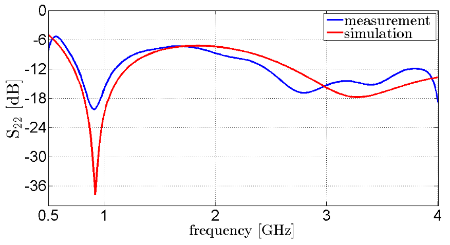
  • The transmission parameter |𝑆12| between a monopole antenna (port 2) vertically placed in the center of the head phantom and a vertically polarized horn antenna (port 1) placed at 1m distance is measured between 0.5 and 4 GHz with a HP 8720D to ensure far field conditions of a linearly polarized plane wave, along the z-direction.
  • This figure shows reasonable agreements between the measured and the simulated data of the same configurations.
  • In the lower part of Fig.8, the product of all these influences gives the total influence 𝑅(𝜔), of all the layers on the wave propagation.
  • It appears clearly that up to around 1.2GHz the “barrier” formed by the CSF, bone, fat and skin, is beneficial to the power transmission, and the opposite above this limit.

IV. CONCLUSION

  • The analysis of the normalized transmitted power of an impinging electromagnetic field onto a simplified model of the human head for MWI applications has been presented using a spherical multilayered model.
  • Additionally, the authors presented a methodology to perform measurements outside and inside liquid phantoms using the 3D printing technology.
  • The strong attenuation of at least 15dB between 1.5GHz and 3GHz in the measurements matches the predictions made with simple transmission line models [14].
  • The experimental results have been compared to theoretical results based on a spherical wave expansion and showed reasonable agreement.
  • This information provides insight on the limit of the frequency, where this parameter starts to have a negative impact on the signal transmission between the brain and the matching medium, and therefore on the imaging quality.

Did you find this useful? Give us your feedback

Content maybe subject to copyright    Report

HAL Id: hal-01739926
https://hal-univ-rennes1.archives-ouvertes.fr/hal-01739926
Submitted on 21 Jan 2021
HAL is a multi-disciplinary open access
archive for the deposit and dissemination of sci-
entic research documents, whether they are pub-
lished or not. The documents may come from
teaching and research institutions in France or
abroad, or from public or private research centers.
L’archive ouverte pluridisciplinaire HAL, est
destinée au dépôt et à la diusion de documents
scientiques de niveau recherche, publiés ou non,
émanant des établissements d’enseignement et de
recherche français ou étrangers, des laboratoires
publics ou privés.
Stratied spherical model for microwave imaging of the
brain Analysis and experimental validation of
transmitted power
M. Bjelogrlic, M. Volery, Benjamin Fuchs, J.-P. Thiran, J.R. Mosig, M. Mattes
To cite this version:
M. Bjelogrlic, M. Volery, Benjamin Fuchs, J.-P. Thiran, J.R. Mosig, et al.. Stratied spherical model
for microwave imaging of the brain Analysis and experimental validation of transmitted power. Mi-
crowave and Optical Technology Letters, Wiley, 2018, 60 (4), pp.1042-1048. �10.1002/mop.31101�.
�hal-01739926�

1
Stratified Spherical Model for Microwave Imaging of the Brain: Analysis and
Experimental Validation of Transmitted Power
Mina Bjelogrlic, Maxime Volery, Benjamin Fuchs, Jean-Philippe Thiran, Juan R. Mosig, and Michael
Mattes
AbstractThis work presents the analysis of power transmission of a radiating field inside the human
head for microwave imaging applications. For this purpose, a spherical layered model composed of
dispersive biological tissues is investigated in the range of [0.5-4] GHz and is confronted to
experimental verification.
Index TermsMicrowave Imaging, bio-medical applications, spherical wave expansion, 3D printed head
phantom.
I. INTRODUCTION
Microwave Imaging (MWI) [1] for bio-medical applications aims at localizing and reconstructing a
pathological tissue region from scattered microwaves. In the framework of head MWI this non-invasive,
non-ionizing technique is suitable to monitor brain anomalies such as brain stroke [2], internal bleeding, etc.
Several main factors influence the quality of the MWI: the scattered power, the frequency and the medium
in which the test object is immersed, called the matching or the background medium. The frequency is an
essential parameter since all biological tissues are dispersive and the losses increase exponentially according
to it.
Another important parameter is the spatial resolution of MWI which has a lower bound defined by the far
field and only depends on the wavelength in the background medium. Devaney [3] first suggested that this
limit of resolution was about
, then Bolomey and Pichot [4] estimated it to
. Chen and Chew [5]
experimentally observed a resolution up to
, for high contrast, but non-dispersive and lossy objects,

2
which is a so-called super-resolution behavior, exploiting the near field and non-linear reconstruction
algorithms. Meaney et al. [6] have suggested that image reconstruction “is fundamentally unlimited by
wavelength” and “is restricted by signal-to-noise” ratio. The latter papers have been experimentally
discussed by Semenov [7] and tested for the detection of myocardial ischemia and infarction. They
concluded that the resolution lies between a quarter and a half of a wavelength in the background medium.
Several groups have developed complex imaging setups and algorithms for imaging brain anomalies [8] [9]
[10] [11]. Experimental phantoms for the head have been developed, the most complex one using molded
semi-rigid parts assembled inside an outer solid cavity [12]. The complex molding procedure and the high
number of used ingredients makes this approach precise, however not easily repeatable. Moreover, the skin
is not represented realistically as it is included in the fat/bone layer printed in a plastic material with
dielectric constant of 6 at 2 GHz [13]. These models are very useful to realistically simulate the dielectric
properties inside the brain itself and are used for imaging [9]. To validate numerical models, interesting
works have been done in the microwave imaging community with a 3D printed breast phantoms [17] [18]
with Triton X-100 based mixtures to mimic biological tissues. In the 7T MRI community 3D printed liquid
phantoms [19] are also used as they are easy to handle and transportable. A similar approach is followed
here, to our best knowledge, for the first time in the framework of MWI of the brain, and a first attempt of
using this simple, repeatable and over time stable procedure was published recently in [20].
In [14] guidelines to design an optimal MWI setup and to properly set the working frequency and the
matching medium, needed to facilitate the penetration of the probing wave into the head, are determined
using a plane wave Transmission Line (TL) model since it allows a simple analysis. On the other hand, a
multilayered spherical model better approximates the head geometry while still allowing an analytical
solution to the electromagnetic scattering problem [15] [16].
This paper focuses on the analysis of the power transmission through the four main layers between the brain
and the background medium, namely the Cerebrospinal Fluid (CSF), the bone, the fat and the skin. These
four layers have very different influence on the propagation of the wave from and into the brain, according

3
to the frequency. For this, a spherically stratified head phantom has been built to experimentally estimate
the power transmitted into the brain and to analyze the influence of these layers on the propagation of the
EM wave.
II. MODEL AND ELECTROMAGNETIC ANALYSIS
Figure 1: Spherical multilayered model to analyze the power transmitted into the human head.
The spherically stratified model, sketched in Fig. 1 (right), is a more realistic model of the human head than
the planar model in [14] while still allowing an analytic solution for the electric field distribution. The core
of the sphere is the brain with a radius of
mm, the

layer (the core is the

layer) is given by

, , and represents, respectively, the CSF (3mm), the cortical bone (7mm) (denoted
here bone), the fat (4mm), the skin (4mm) and the matching medium (see Fig. 1). In [19] for 7T MRI the
Larmor frequency is around 300MHz and the brain region is modeled as a combination of CSF, grey matter,
and white matter. Since we deal with higher frequencies and, thus, shorter wavelengths, we model the CSF
separately and the dielectric characteristics of the brain are set with a grey over white matter ratio (GM/WM

4
ratio) of 1.5 as shown in [21]. A similar ratio was used in [20], as the dielectric properties of the latter two
are similar in the frequency range of interest.
Due to the spherical geometry of the boundary conditions, the electric field can be expanded as an infinite
sum of vector spherical harmonics and be expressed analytically. Reference [22] provides a review on the
governing equations of the spherical wave expansion used to solve this problem, and gives a detailed
analysis and validations of the implementation. These results are valid for a plane wave impinging on the
head phantom. This needs to be taken into account when comparing theoretical and experimental results.
However, because we are only interested in the transmission inside the head, this is not a real restriction and
the results using a plane wave should be also valid for an antenna directly placed on the head since the
propagation of an electromagnetic wave depends only on the properties of the medium and not on the
characteristics of the wave, that is plane wave, spherical wave, etc. Finally, we define the normalized
transmitted power,

, as the ratio between the transmitted power into a bounded domain in the center of
the brain and the power available in the lossless background in the very same bounded domain.
C. Simulation Results and Discussion
Figure 2: Normalized transmitted power according to the frequency 󰇟 󰇠GHz, for a matching medium of

󰇟 󰇠
(left). Cuts for

 of the

(right).
The normalized transmitted power represents the incoming power that can excite any anomaly in the center
of the head (worst case scenario) and therefore produce a scattered field. Fig. 2 represents a map of the

Citations
More filters
Journal ArticleDOI
18 Dec 2018
TL;DR: It is shown herein that breast and head phantoms fabricated from 3D-printed structures and liquid mixtures can also accurately mimic most of the head tissues and that, given a binary fluid mixture model, the respective concentrations of the various constituents needed to mimic a particular tissue can be predetermined by means of a standard minimization method.
Abstract: This paper deals with breast and head phantoms fabricated from 3D-printed structures and liquid mixtures whose complex permittivities are close to that of the biological tissues within a large frequency band. The goal is to enable an easy and safe manufacturing of stable-in-time detailed anthropomorphic phantoms dedicated to the test of microwave imaging systems to assess the performances of the latter in realistic configurations before a possible clinical application to breast cancer imaging or brain stroke monitoring. The structure of the breast phantom has already been used by several laboratories to test their measurement systems in the framework of the COST (European Cooperation in Science and Technology) Action TD1301-MiMed. As for the tissue mimicking liquid mixtures, they are based upon Triton X-100 and salted water. It has been proven that such mixtures can dielectrically mimic the various breast tissues. It is shown herein that they can also accurately mimic most of the head tissues and that, given a binary fluid mixture model, the respective concentrations of the various constituents needed to mimic a particular tissue can be predetermined by means of a standard minimization method.

66 citations

Journal ArticleDOI
TL;DR: A microwave tomographic approach for the quantitative imaging of brain stroke inside the human head using a prototype of multistatic system based on a variable-exponent Lebesgue-space regularization technique, whose outcome is a map of dielectric properties of a slice of the head.
Abstract: This article describes a microwave tomographic approach for the quantitative imaging of brain stroke inside the human head. For the acquisition of the scattered-field information, a prototype of multistatic system is adopted. An array of custom antennas is placed in contact with the head, and a switching matrix is used to measure the scattering parameters for each pair of probes. The collected data are processed by an inversion method based on a variable-exponent Lebesgue-space regularization technique, whose outcome is a map of dielectric properties of a slice of the head. With respect to previous approaches, this kind of inversion procedure performs an adaptive update of the Lebesgue-space exponents on the basis of the results at each inexact-Newton iteration and exploits stepped frequency data. This allows for an automatic setting of the regularization level, which becomes variable and target-dependent inside the whole investigation domain. The proposed approach is validated by means of FDTD synthetic simulations with a realistic 3-D forward scattering model of the human head, as well as by using real experimental cylindrical test phantoms filled with saline and glycerin/water mixtures.

49 citations


Additional excerpts

  • ..., elliptical and spherical layered models [59]...

    [...]

Journal ArticleDOI
09 Aug 2019-Sensors
TL;DR: Simulated microwave transmission data is used to investigate the performance of a machine learning classification algorithm based on subspace distances for the detection of intracranial bleeding and shows that classification results improved with the number of subjects in the training data.
Abstract: Early, preferably prehospital, detection of intracranial bleeding after trauma or stroke would dramatically improve the acute care of these large patient groups. In this paper, we use simulated microwave transmission data to investigate the performance of a machine learning classification algorithm based on subspace distances for the detection of intracranial bleeding. A computational model, consisting of realistic human head models of patients with bleeding, as well as healthy subjects, was inserted in an antenna array model. The Finite-Difference Time-Domain (FDTD) method was then used to generate simulated transmission coefficients between all possible combinations of antenna pairs. These transmission data were used both to train and evaluate the performance of the classification algorithm and to investigate its ability to distinguish patients with versus without intracranial bleeding. We studied how classification results were affected by the number of healthy subjects and patients used to train the algorithm, and in particular, we were interested in investigating how many samples were needed in the training dataset to obtain classification results better than chance. Our results indicated that at least 200 subjects, i.e., 100 each of the healthy subjects and bleeding patients, were needed to obtain classification results consistently better than chance (p < 0.05 using Student's t-test). The results also showed that classification results improved with the number of subjects in the training data. With a sample size that approached 1000 subjects, classifications results characterized as area under the receiver operating curve (AUC) approached 1.0, indicating very high sensitivity and specificity.

12 citations


Cites background from "Stratified spherical model for micr..."

  • ...Significant efforts, including experimental lab test on phantoms, were published in [11]....

    [...]

Journal ArticleDOI
TL;DR: The experimental results show that the system for identifying cerebral stroke based on microwaves can distinguish between cerebral hemorrhage and cerebral ischemia models and effectively distinguish between different degrees of cerebral hemorrhages or different durations of cerebral ischemic stroke.
Abstract: Background Hemorrhagic stroke and ischemic stroke have similar symptoms at the onset of the disease, but their clinical treatment is completely different. The early, effective identification of stroke types can effectively improve the cure rate. Objective In this study, an early, noncontact identification of the stroke type, i.e., hemorrhagic or ischemic, based on a microwave measurement technique was investigated. Methods This study was based on animal models of cerebral hemorrhage and cerebral ischemia and the design of a microwave scattering parameter measurement system. Results The accuracy of the cerebral hemorrhage model with a blood loss interval of 2 ml reached 93.75%. While the accuracy of the cerebral ischemia model with an ischemic interval of 42 minutes reached 91.7%. Conclusion The experimental results show that the system for identifying cerebral stroke based on microwaves can distinguish between cerebral hemorrhage and cerebral ischemia models and effectively distinguish between different degrees of cerebral hemorrhage or different durations of cerebral ischemia. This experimental system is inexpensive, portable, noninvasive, simple, and rapid and thus has good potential as a method for identifying the stroke type prior to hospitalization.

8 citations

Journal ArticleDOI
22 Feb 2021
TL;DR: In this article, a general methodology for the development of a biological head phantom is presented, and this approach is applied to the particular case of the experimental device developed by the Department of Electronics and Telecommunications at Politecnico di Torino (POLITO) that currently uses a homogeneous version of the head phantom considered in this paper.
Abstract: This work is devoted to the development and manufacturing of realistic benchmark phantoms to evaluate the performance of microwave imaging devices. The 3D (3 dimensional) printed phantoms contain several cavities, designed to be filled with liquid solutions that mimic biological tissues in terms of complex permittivity over a wide frequency range. Numerical versions (stereolithography (STL) format files) of these phantoms were used to perform simulations to investigate experimental parameters. The purpose of this paper is two-fold. First, a general methodology for the development of a biological phantom is presented. Second, this approach is applied to the particular case of the experimental device developed by the Department of Electronics and Telecommunications at Politecnico di Torino (POLITO) that currently uses a homogeneous version of the head phantom considered in this paper. Numerical versions of the introduced inhomogeneous head phantoms were used to evaluate the effect of various parameters related to their development, such as the permittivity of the equivalent biological tissue, coupling medium, thickness and nature of the phantom walls, and number of compartments. To shed light on the effects of blood circulation on the recognition of a randomly shaped stroke, a numerical brain model including blood vessels was considered.

4 citations

References
More filters
Journal ArticleDOI
TL;DR: In this article, the dielectric properties of various mixtures susceptible to be used in manufacturing of reference inhomogeneous breast phantoms dedicated to the experimental validation of microwave breast imaging systems in the 0.5-6 GHz frequency range were investigated.
Abstract: Herein, we study the dielectric properties of various mixtures susceptible to be used in manufacturing of reference inhomogeneous breast phantoms dedicated to the experimental validation of microwave breast imaging systems in the 0.5-6-GHz frequency range. Particularly, we investigate the stability over time and temperature of these properties and their reproducibility for a given mixture, as well as the ability of some mixtures to mimic the various breast tissues, i.e., to show dielectric properties close to that given by one-pole Debye models that describe the mean relative dielectric permittivity of various tissue types.

92 citations

Journal ArticleDOI
TL;DR: This paper presents Debye potential (DP) and Dyadic Green's function (DGF)-based solutions of the EMFs inside a head-sized, stratified sphere with similar radial conductivity and permittivity profiles as a human head for rapid prototyping.
Abstract: The precise evaluation of electromagnetic field (EMF) distributions inside biological samples is becoming an increasingly important design requirement for high field MRI systems. In evaluating the induced fields caused by magnetic field gradients and RF transmitter coils, a multilayered dielectric spherical head model is proposed to provide a better understanding of electromagnetic interactions when compared to a traditional homogeneous head phantom. This paper presents Debye potential (DP) and Dyadic Green's function (DGF)-based solutions of the EMFs inside a head-sized, stratified sphere with similar radial conductivity and permittivity profiles as a human head. The DP approach is formulated for the symmetric case in which the source is a circular loop carrying a harmonic-formed current over a wide frequency range. The DGF method is developed for generic cases in which the source may be any kind of RF coil whose current distribution can be evaluated using the method of moments. The calculated EMFs can then be used to deduce MRI imaging parameters. The proposed methods, while not representing the full complexity of a head model, offer advantages in rapid prototyping as the computation times are much lower than a full finite difference time domain calculation using a complex head model. Test examples demonstrate the capability of the proposed models/methods. It is anticipated that this model will be of particular value for high field MRI applications, especially the rapid evaluation of RF resonator (surface and volume coils) and high performance gradient set designs.

85 citations

Journal ArticleDOI
TL;DR: It has been found that formulating the imaging algorithm in terms of the logarithm of the amplitude and the unwrapped phase of the measured signals improves its performance when compared to the more commonly used complex phasor formulation.
Abstract: The microwave imaging system currently being developed at the Technical University of Denmark is described and its performance tested on simulated data. The system uses an iterative Newton-based imaging algorithm for reconstructing the images in conjunction with an efficient method-of-moments solution of the associated forward scattering problem. A cylindrical multistatic antenna setup with 32 horizontally oriented antennas is used for collecting the data. It has been found that formulating the imaging algorithm in terms of the logarithm of the amplitude and the unwrapped phase of the measured signals improves its performance when compared to the more commonly used complex phasor formulation. This improvement is illustrated by imaging a simulated hemispherical breast model using both formulations. In addition to this, the importance of using the correct position and orientation of the antennas in the measurement system is shown by imaging the same breast model using a measurement setup in which the antennas are vertically oriented.

85 citations

Journal ArticleDOI
TL;DR: In this paper, the authors presented an experimental verification of the super-resolution phenomenon in a nonlinear inverse scattering algorithm, namely the distorted Born iterative method, by using an experimental setup based on a recently developed time-domain ultra-wide-band radar imaging system at the University of Illinois at Urbana-Champaign.
Abstract: This letter presents an experimental verification of the super-resolution phenomenon in a nonlinear inverse scattering algorithm, namely the distorted Born iterative method, by using an experimental setup based on a recently developed time-domain ultra-wide-band radar imaging system at the University of Illinois at Urbana-Champaign. The experimental result also demonstrates that the distorted Born iterative method can recover information on the backside of a penetrable scatterer even though scattering data are collected only from limited viewing angles on the front side of the scatterer.

82 citations

Journal ArticleDOI
13 Apr 2016-PLOS ONE
TL;DR: The proposed head imaging prototype along with the processing algorithm demonstrates its feasibility for potential use in ambulances as an effective and low cost diagnostic tool to assure timely triaging of intracranial hemorrhage patients.
Abstract: An intracranial haemorrhage is a life threatening medical emergency, yet only a fraction of the patients receive treatment in time, primarily due to the transport delay in accessing diagnostic equipment in hospitals such as Magnetic Resonance Imaging or Computed Tomography. A mono-static microwave head imaging system that can be carried in an ambulance for the detection and localization of intracranial haemorrhage is presented. The system employs a single ultra-wideband antenna as sensing element to transmit signals in low microwave frequencies towards the head and capture backscattered signals. The compact and low-profile antenna provides stable directional radiation patterns over the operating bandwidth in both near and far-fields. Numerical analysis of the head imaging system with a realistic head model in various situations is performed to realize the scattering mechanism of haemorrhage. A modified delay-and-summation back-projection algorithm, which includes effects of surface waves and a distance-dependent effective permittivity model, is proposed for signal and image post-processing. The efficacy of the automated head imaging system is evaluated using a 3D-printed human head phantom with frequency dispersive dielectric properties including emulated haemorrhages with different sizes located at different depths. Scattered signals are acquired with a compact transceiver in a mono-static circular scanning profile. The reconstructed images demonstrate that the system is capable of detecting haemorrhages as small as 1 cm3. While quantitative analyses reveal that the quality of images gradually degrades with the increase of the haemorrhage’s depth due to the reduction of signal penetration inside the head; rigorous statistical analysis suggests that substantial improvement in image quality can be obtained by increasing the data samples collected around the head. The proposed head imaging prototype along with the processing algorithm demonstrates its feasibility for potential use in ambulances as an effective and low cost diagnostic tool to assure timely triaging of intracranial hemorrhage patients.

71 citations

Frequently Asked Questions (14)
Q1. What are the contributions mentioned in the paper "Stratified spherical model for microwave imaging of the brain analysis and experimental validation of transmitted power" ?

This work presents the analysis of power transmission of a radiating field inside the human head for microwave imaging applications. 

Due to the spherical geometry of the boundary conditions, the electric field can be expanded as an infinite sum of vector spherical harmonics and be expressed analytically. 

At 1GHz for example, the optimum is at 𝜀𝑚𝑚 = 10, however the normalized transmitted power drops by approximately 3dB if 𝜀𝑚𝑚 = 80 (approximately water at 1GHz), but the imaging resolution would increase by almost a factor of 3. 

Depending on the sensitivity of the data acquisition of the imaging system, the frequency and matching medium ranges can be chosen using simplified analytical models and then be fine-tuned using more complex EM solvers and more realistic models of the head. 

GHz the normalized transmitted power 𝑃𝑁𝑡 drops very rapidly by 15dB due to the strong attenuation in the tissues, which was predicted by both the planar and the spherical model. 

In [19] for 7T MRI the Larmor frequency is around 300MHz and the brain region is modeled as a combination of CSF, grey matter, and white matter. 

The transmission parameter |𝑆12| between a monopole antenna (port 2) vertically placed in the center of the head phantom and a vertically polarized horn antenna (port 1) placed at 1m distance is measured between 0.5 and 4 GHz with a HP 8720D to ensure far field conditions of a linearly polarized plane wave, along the z-direction. 

The dielectric properties of the ABS plastic structure of the 3D printed prototype were measured in the range of [0.5 − 4] GHz using the Agilent 85070E dielectric probe kit. 

The color change corresponds to a drop in the normalized transmitted power in steps of 3dB and up to -36dB (all values below -36dB are depicted as the same dark blue color). 

The almost free choice of the permittivity (1dB drop of 𝑃𝑁𝑡 for increasing 𝜀𝑚𝑚 from 56 to 80) means also, that it can be used to improve the imaging resolution according to the discussion in the introduction. 

The strong attenuation of at least 15dB between 1.5GHz and 3GHz in the measurements matches the predictions made with simple transmission line models [14]. 

As the filling process allowed to fill each layer on-site without moving the prototype (see Fig. 5), it was possible to estimate the influence of each layer on the power transmission. 

because the authors are only interested in the transmission inside the head, this is not a real restriction and the results using a plane wave should be also valid for an antenna directly placed on the head since the propagation of an electromagnetic wave depends only on the properties of the medium and not on the characteristics of the wave, that is plane wave, spherical wave, etc. 

The spherically stratified model, sketched in Fig. 1 (right), is a more realistic model of the human head than the planar model in [14] while still allowing an analytic solution for the electric field distribution.