Example of Oral Radiology format
Recent searches

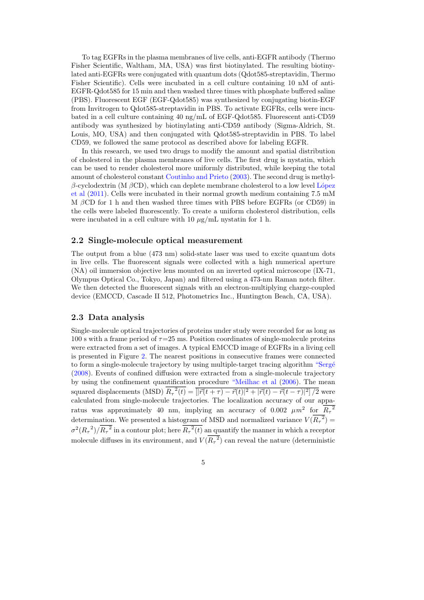
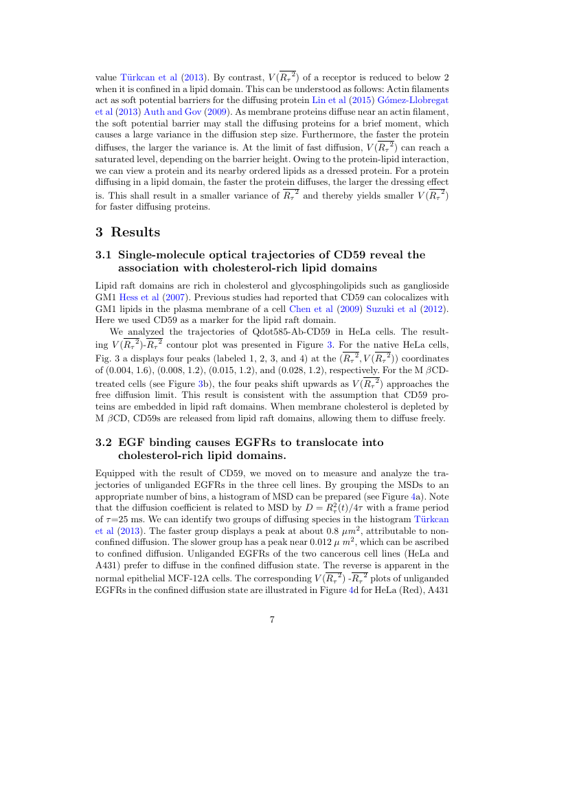
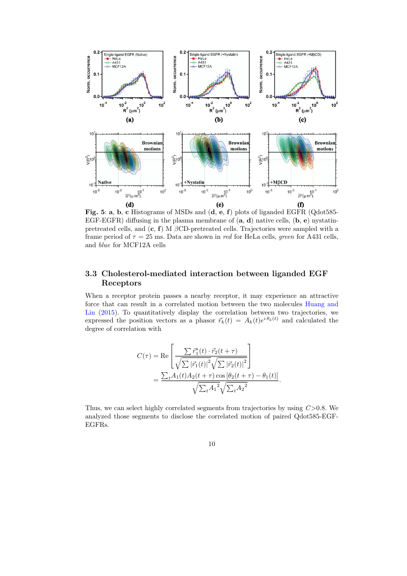
Example of Oral Radiology format Example of Oral Radiology format Example of Oral Radiology format Example of Oral Radiology format Example of Oral Radiology format Example of Oral Radiology format Example of Oral Radiology format Example of Oral Radiology format Example of Oral Radiology format Example of Oral Radiology format Example of Oral Radiology format Example of Oral Radiology format Example of Oral Radiology format Example of Oral Radiology format Example of Oral Radiology format Example of Oral Radiology format Example of Oral Radiology format Example of Oral Radiology format
Sample paper formatted on SciSpace - SciSpace
This content is only for preview purposes. The original open access content can be found here.
Look Inside
Example of Oral Radiology format Example of Oral Radiology format Example of Oral Radiology format Example of Oral Radiology format Example of Oral Radiology format Example of Oral Radiology format Example of Oral Radiology format Example of Oral Radiology format Example of Oral Radiology format Example of Oral Radiology format Example of Oral Radiology format Example of Oral Radiology format Example of Oral Radiology format Example of Oral Radiology format Example of Oral Radiology format Example of Oral Radiology format Example of Oral Radiology format Example of Oral Radiology format
Sample paper formatted on SciSpace - SciSpace
This content is only for preview purposes. The original open access content can be found here.
open access Open Access

Oral Radiology — Template for authors

Publisher: Springer
Categories Rank Trend in last 3 yrs
Dentistry (miscellaneous) #9 of 16 up up by 2 ranks
Radiology, Nuclear Medicine and Imaging #198 of 288 up up by 12 ranks
journal-quality-icon Journal quality:
Medium
calendar-icon Last 4 years overview: 173 Published Papers | 282 Citations
indexed-in-icon Indexed in: Scopus
last-updated-icon Last updated: 02/06/2020
Related journals
Insights
General info
Top papers
Popular templates
Get started guide
Why choose from SciSpace
FAQ

Related Journals

open access Open Access

Elsevier

Quality:  
High
CiteRatio: 3.1
SJR: 0.664
SNIP: 1.265
open access Open Access
recommended Recommended

SAGE

Quality:  
High
CiteRatio: 5.5
SJR: 1.72
SNIP: 1.405
open access Open Access
recommended Recommended

Hindawi

Quality:  
High
CiteRatio: 7.9
SJR: 0.626
SNIP: 3.203
open access Open Access

SPIE

Quality:  
High
CiteRatio: 4.7
SJR: 0.779
SNIP: 1.165

Journal Performance & Insights

Impact Factor

CiteRatio

Determines the importance of a journal by taking a measure of frequency with which the average article in a journal has been cited in a particular year.

A measure of average citations received per peer-reviewed paper published in the journal.

0.54

21% from 2018

Impact factor for Oral Radiology from 2016 - 2019
Year Value
2019 0.54
2018 0.681
2017 0.466
2016 0.554
graph view Graph view
table view Table view

1.6

78% from 2019

CiteRatio for Oral Radiology from 2016 - 2020
Year Value
2020 1.6
2019 0.9
2018 0.8
2017 1.0
2016 0.9
graph view Graph view
table view Table view

insights Insights

  • Impact factor of this journal has decreased by 21% in last year.
  • This journal’s impact factor is in the top 10 percentile category.

insights Insights

  • CiteRatio of this journal has increased by 78% in last years.
  • This journal’s CiteRatio is in the top 10 percentile category.

SCImago Journal Rank (SJR)

Source Normalized Impact per Paper (SNIP)

Measures weighted citations received by the journal. Citation weighting depends on the categories and prestige of the citing journal.

Measures actual citations received relative to citations expected for the journal's category.

0.434

113% from 2019

SJR for Oral Radiology from 2016 - 2020
Year Value
2020 0.434
2019 0.204
2018 0.277
2017 0.246
2016 0.187
graph view Graph view
table view Table view

0.74

91% from 2019

SNIP for Oral Radiology from 2016 - 2020
Year Value
2020 0.74
2019 0.388
2018 0.412
2017 0.356
2016 0.432
graph view Graph view
table view Table view

insights Insights

  • SJR of this journal has increased by 113% in last years.
  • This journal’s SJR is in the top 10 percentile category.

insights Insights

  • SNIP of this journal has increased by 91% in last years.
  • This journal’s SNIP is in the top 10 percentile category.

Oral Radiology

Guideline source: View

All company, product and service names used in this website are for identification purposes only. All product names, trademarks and registered trademarks are property of their respective owners.

Use of these names, trademarks and brands does not imply endorsement or affiliation. Disclaimer Notice

Springer

Oral Radiology

As the official English-language journal of the Japanese Society for Oral and Maxillofacial Radiology, Oral Radiology is intended to be a forum for international collaboration in head and neck diagnostic imaging and all related fields. Oral Radiology features cutting-edge rese...... Read More

Dentistry

i
Last updated on
02 Jun 2020
i
ISSN
0911-6028
i
Impact Factor
Low - 0.337
i
Open Access
No
i
Sherpa RoMEO Archiving Policy
Green faq
i
Plagiarism Check
Available via Turnitin
i
Endnote Style
Download Available
i
Bibliography Name
SPBASIC
i
Citation Type
Author Year
(Blonder et al, 1982)
i
Bibliography Example
Beenakker CWJ (2006) Specular andreev reflection in graphene. Phys Rev Lett 97(6):067,007, URL 10.1103/PhysRevLett.97.067007

Top papers written in this journal

Journal Article DOI: 10.1007/S11282-018-0363-7
Deep-learning classification using convolutional neural network for evaluation of maxillary sinusitis on panoramic radiography
01 Sep 2019 - Oral Radiology

Abstract:

To apply a deep-learning system for diagnosis of maxillary sinusitis on panoramic radiography, and to clarify its diagnostic performance. Training data for 400 healthy and 400 inflamed maxillary sinuses were enhanced to 6000 samples in each category by data augmentation. Image patches were input into a deep-learning system, t... To apply a deep-learning system for diagnosis of maxillary sinusitis on panoramic radiography, and to clarify its diagnostic performance. Training data for 400 healthy and 400 inflamed maxillary sinuses were enhanced to 6000 samples in each category by data augmentation. Image patches were input into a deep-learning system, the learning process was repeated for 200 epochs, and a learning model was created. Newly-prepared testing image patches from 60 healthy and 60 inflamed sinuses were input into the learning model, and the diagnostic performance was calculated. Receiver-operating characteristic (ROC) curves were drawn, and the area under the curve (AUC) values were obtained. The results were compared with those of two experienced radiologists and two dental residents. The diagnostic performance of the deep-learning system for maxillary sinusitis on panoramic radiographs was high, with accuracy of 87.5%, sensitivity of 86.7%, specificity of 88.3%, and AUC of 0.875. These values showed no significant differences compared with those of the radiologists and were higher than those of the dental residents. The diagnostic performance of the deep-learning system for maxillary sinusitis on panoramic radiographs was sufficiently high. Results from the deep-learning system are expected to provide diagnostic support for inexperienced dentists. read more read less
114 Citations
Journal Article DOI: 10.1007/S11282-019-00409-X
Evaluation of an artificial intelligence system for detecting vertical root fracture on panoramic radiography
01 Oct 2020 - Oral Radiology

Abstract:

The aim of this study was to evaluate the use of a convolutional neural network (CNN) system for detecting vertical root fracture (VRF) on panoramic radiography. Three hundred panoramic images containing a total of 330 VRF teeth with clearly visible fracture lines were selected from our hospital imaging database. Confirmation... The aim of this study was to evaluate the use of a convolutional neural network (CNN) system for detecting vertical root fracture (VRF) on panoramic radiography. Three hundred panoramic images containing a total of 330 VRF teeth with clearly visible fracture lines were selected from our hospital imaging database. Confirmation of VRF lines was performed by two radiologists and one endodontist. Eighty percent (240 images) of the 300 images were assigned to a training set and 20% (60 images) to a test set. A CNN-based deep learning model for the detection of VRFs was built using DetectNet with DIGITS version 5.0. To defend test data selection bias and increase reliability, fivefold cross-validation was performed. Diagnostic performance was evaluated using recall, precision, and F measure. Of the 330 VRFs, 267 were detected. Twenty teeth without fractures were falsely detected. Recall was 0.75, precision 0.93, and F measure 0.83. The CNN learning model has shown promise as a tool to detect VRFs on panoramic images and to function as a CAD tool. read more read less
106 Citations
Journal Article DOI: 10.1007/S11282-011-0074-9
Observation of the retromolar foramen and canal of the mandible: a CBCT and macroscopic study
Taisuke Kawai1, Rieko Asaumi1, Iwao Sato, Yasuo Kumazawa, Takashi Yosue1
01 Mar 2012 - Oral Radiology

Abstract:

The retromolar foramen (RMF) is an anatomical structure on the alveolar surface of the retromolar area. This foramen runs consecutive to the retromolar canal (RMC), which diverges from the mandibular canal. It is important to confirm the RMF and canal locations prior to surgical procedures, such as extraction of an impacted m... The retromolar foramen (RMF) is an anatomical structure on the alveolar surface of the retromolar area. This foramen runs consecutive to the retromolar canal (RMC), which diverges from the mandibular canal. It is important to confirm the RMF and canal locations prior to surgical procedures, such as extraction of an impacted molar and bone harvesting as a donor site for bone graft surgery. This aim of this study was to investigate the RMF in Japanese cadaver mandibles using cone-beam computed tomography (CBCT) images and anatomical observations. Ninety sides of 46 cadaver mandibles were investigated in this study. CBCT images around the retromolar region were acquired for all of the mandibles. The frequency and anteroposterior and buccolingual locations of the RMF were examined on these images. Subsequently, four sides of three mandibles were dissected to confirm the contents of the RMC/RMF. In 24 of 46 (52%) mandibles and 34 of 90 (37%) sides, at least one RMF was observed in the images. In 26 dentate mandibles, 12 (48%) mandibles and 14 (33%) sides presented at least one RMF. The average location of the RMF was 14.4 mm posterior from the distal edge of the second molar. The buccolingual location was 3.0 mm lingual from the mandibular canal. Observations made during the cadaver dissections confirmed that the vessels and nerves diverged from the mandibular canal. The findings suggest that the RMF is not a rare anatomical structure and that practitioners should take this foramen into account in all anesthetic and surgical procedures involving the retromolar area. read more read less

Topics:

Mandibular canal (59%)59% related to the paper, Mandibular second molar (51%)51% related to the paper, Foramen (51%)51% related to the paper
59 Citations
Journal Article DOI: 10.1007/S11282-011-0071-Z
Quantitative analysis of metallic artifacts caused by dental metals: comparison of cone-beam and multi-detector row CT scanners
30 Jun 2011 - Oral Radiology

Abstract:

To quantitatively compare the streak artifacts produced by dental metals in a cone-beam computed tomography (CBCT) device and a multi-detector row computed tomography (MDCT) scanner in relation to metal types and imaging parameters. Cubes of aluminum, titanium, cobalt–chromium alloy, and type IV gold alloy were scanned with C... To quantitatively compare the streak artifacts produced by dental metals in a cone-beam computed tomography (CBCT) device and a multi-detector row computed tomography (MDCT) scanner in relation to metal types and imaging parameters. Cubes of aluminum, titanium, cobalt–chromium alloy, and type IV gold alloy were scanned with CBCT and MDCT scanners at tube voltages of 80 and 100 peak kV (kVp), and currents of 100 and 170 mAs by MDCT, and 102 and 170 mAs by CBCT. Artifact areas were quantified using ImageJ software. Artifact areas for the same metals and imaging parameters were smaller with CBCT than with MDCT under most conditions. Type IV gold alloy caused the largest artifact areas, followed by cobalt–chromium alloy, titanium, and aluminum, respectively. Higher tube voltage was associated with smaller artifact areas under most conditions, whereas increasing tube current had no consistent effect on artifact area using either CT device. CBCT was associated with smaller artifact areas than MDCT for the same parameters. Type IV gold alloy produced the largest artifact areas among the tested metals, but metallic artifacts could be reduced by increasing the tube voltage. read more read less

Topics:

Cone beam computed tomography (56%)56% related to the paper, Artifact (error) (54%)54% related to the paper
58 Citations
Author Pic

SciSpace is a very innovative solution to the formatting problem and existing providers, such as Mendeley or Word did not really evolve in recent years.

- Andreas Frutiger, Researcher, ETH Zurich, Institute for Biomedical Engineering

Get MS-Word and LaTeX output to any Journal within seconds
1
Choose a template
Select a template from a library of 40,000+ templates
2
Import a MS-Word file or start fresh
It takes only few seconds to import
3
View and edit your final output
SciSpace will automatically format your output to meet journal guidelines
4
Submit directly or Download
Submit to journal directly or Download in PDF, MS Word or LaTeX

(Before submission check for plagiarism via Turnitin)

clock Less than 3 minutes

What to expect from SciSpace?

Speed and accuracy over MS Word

''

With SciSpace, you do not need a word template for Oral Radiology.

It automatically formats your research paper to Springer formatting guidelines and citation style.

You can download a submission ready research paper in pdf, LaTeX and docx formats.

Time comparison

Time taken to format a paper and Compliance with guidelines

Plagiarism Reports via Turnitin

SciSpace has partnered with Turnitin, the leading provider of Plagiarism Check software.

Using this service, researchers can compare submissions against more than 170 million scholarly articles, a database of 70+ billion current and archived web pages. How Turnitin Integration works?

Turnitin Stats
Publisher Logos

Freedom from formatting guidelines

One editor, 100K journal formats – world's largest collection of journal templates

With such a huge verified library, what you need is already there.

publisher-logos

Easy support from all your favorite tools

Oral Radiology format uses SPBASIC citation style.

Automatically format and order your citations and bibliography in a click.

SciSpace allows imports from all reference managers like Mendeley, Zotero, Endnote, Google Scholar etc.

Frequently asked questions

1. Can I write Oral Radiology in LaTeX?

Absolutely not! Our tool has been designed to help you focus on writing. You can write your entire paper as per the Oral Radiology guidelines and auto format it.

2. Do you follow the Oral Radiology guidelines?

Yes, the template is compliant with the Oral Radiology guidelines. Our experts at SciSpace ensure that. If there are any changes to the journal's guidelines, we'll change our algorithm accordingly.

3. Can I cite my article in multiple styles in Oral Radiology?

Of course! We support all the top citation styles, such as APA style, MLA style, Vancouver style, Harvard style, and Chicago style. For example, when you write your paper and hit autoformat, our system will automatically update your article as per the Oral Radiology citation style.

4. Can I use the Oral Radiology templates for free?

Sign up for our free trial, and you'll be able to use all our features for seven days. You'll see how helpful they are and how inexpensive they are compared to other options, Especially for Oral Radiology.

5. Can I use a manuscript in Oral Radiology that I have written in MS Word?

Yes. You can choose the right template, copy-paste the contents from the word document, and click on auto-format. Once you're done, you'll have a publish-ready paper Oral Radiology that you can download at the end.

6. How long does it usually take you to format my papers in Oral Radiology?

It only takes a matter of seconds to edit your manuscript. Besides that, our intuitive editor saves you from writing and formatting it in Oral Radiology.

7. Where can I find the template for the Oral Radiology?

It is possible to find the Word template for any journal on Google. However, why use a template when you can write your entire manuscript on SciSpace , auto format it as per Oral Radiology's guidelines and download the same in Word, PDF and LaTeX formats? Give us a try!.

8. Can I reformat my paper to fit the Oral Radiology's guidelines?

Of course! You can do this using our intuitive editor. It's very easy. If you need help, our support team is always ready to assist you.

9. Oral Radiology an online tool or is there a desktop version?

SciSpace's Oral Radiology is currently available as an online tool. We're developing a desktop version, too. You can request (or upvote) any features that you think would be helpful for you and other researchers in the "feature request" section of your account once you've signed up with us.

10. I cannot find my template in your gallery. Can you create it for me like Oral Radiology?

Sure. You can request any template and we'll have it setup within a few days. You can find the request box in Journal Gallery on the right side bar under the heading, "Couldn't find the format you were looking for like Oral Radiology?”

11. What is the output that I would get after using Oral Radiology?

After writing your paper autoformatting in Oral Radiology, you can download it in multiple formats, viz., PDF, Docx, and LaTeX.

12. Is Oral Radiology's impact factor high enough that I should try publishing my article there?

To be honest, the answer is no. The impact factor is one of the many elements that determine the quality of a journal. Few of these factors include review board, rejection rates, frequency of inclusion in indexes, and Eigenfactor. You need to assess all these factors before you make your final call.

13. What is Sherpa RoMEO Archiving Policy for Oral Radiology?

SHERPA/RoMEO Database

We extracted this data from Sherpa Romeo to help researchers understand the access level of this journal in accordance with the Sherpa Romeo Archiving Policy for Oral Radiology. The table below indicates the level of access a journal has as per Sherpa Romeo's archiving policy.

RoMEO Colour Archiving policy
Green Can archive pre-print and post-print or publisher's version/PDF
Blue Can archive post-print (ie final draft post-refereeing) or publisher's version/PDF
Yellow Can archive pre-print (ie pre-refereeing)
White Archiving not formally supported
FYI:
  1. Pre-prints as being the version of the paper before peer review and
  2. Post-prints as being the version of the paper after peer-review, with revisions having been made.

14. What are the most common citation types In Oral Radiology?

The 5 most common citation types in order of usage for Oral Radiology are:.

S. No. Citation Style Type
1. Author Year
2. Numbered
3. Numbered (Superscripted)
4. Author Year (Cited Pages)
5. Footnote

15. How do I submit my article to the Oral Radiology?

It is possible to find the Word template for any journal on Google. However, why use a template when you can write your entire manuscript on SciSpace , auto format it as per Oral Radiology's guidelines and download the same in Word, PDF and LaTeX formats? Give us a try!.

16. Can I download Oral Radiology in Endnote format?

Yes, SciSpace provides this functionality. After signing up, you would need to import your existing references from Word or Bib file to SciSpace. Then SciSpace would allow you to download your references in Oral Radiology Endnote style according to Elsevier guidelines.

Fast and reliable,
built for complaince.

Instant formatting to 100% publisher guidelines on - SciSpace.

Available only on desktops 🖥

No word template required

Typset automatically formats your research paper to Oral Radiology formatting guidelines and citation style.

Verifed journal formats

One editor, 100K journal formats.
With the largest collection of verified journal formats, what you need is already there.

Trusted by academicians

I spent hours with MS word for reformatting. It was frustrating - plain and simple. With SciSpace, I can draft my manuscripts and once it is finished I can just submit. In case, I have to submit to another journal it is really just a button click instead of an afternoon of reformatting.

Andreas Frutiger
Researcher & Ex MS Word user
Use this template