scispace - formally typeset
Open AccessJournal ArticleDOI

The adult scoliosis

Max Aebi
- 18 Nov 2005 - 
- Vol. 14, Iss: 10, pp 925-948
TLDR
Overall, a satisfactory outcome can be expected in well-differentiated indications and properly tailored surgical procedures, although until today prospective, controlled studies with outcome measures and pre- and post-operative patient’s health status are lacking.
Abstract
Adult scoliosis is defined as a spinal deformity in a skeletally mature patient with a Cobb angle of more than 10 degrees in the coronal plain. Adult scoliosis can be separated into four major groups: Type 1: Primary degenerative scoliosis, mostly on the basis of a disc and/or facet joint arthritis, affecting those structures asymmetrically with predominantly back pain symptoms, often accompanied either by signs of spinal stenosis (central as well as lateral stenosis) or without. These curves are often classified as "de novo" scoliosis. Type 2: Idiopathic adolescent scoliosis of the thoracic and/or lumbar spine which progresses in adult life and is usually combined with secondary degeneration and/or imbalance. Some patients had either no surgical treatment or a surgical correction and fusion in adolescence in either the thoracic or thoracolumbar spine. Those patients may develop secondary degeneration and progression of the adjacent curve; in this case those curves belong to the type 3a. Type 3: Secondary adult curves: (a) In the context of an oblique pelvis, for instance, due to a leg length discrepancy or hip pathology or as a secondary curve in idiopathic, neuromuscular and congenital scoliosis, or asymmetrical anomalies at the lumbosacral junction; (b) In the context of a metabolic bone disease (mostly osteoporosis) combined with asymmetric arthritic disease and/or vertebral fractures. Sometimes it is difficult to decide, what exactly the primary cause of the curve was, once it has significantly progressed. However, once an asymmetric load or degeneration occurs, the pathomorphology and pathomechanism in adult scoliosis predominantly located in the lumbar or thoracolumbar spine is quite predictable. Asymmetric degeneration leads to increased asymmetric load and therefore to a progression of the degeneration and deformity, as either scoliosis and/or kyphosis. The progression of a curve is further supported by osteoporosis, particularly in post-menopausal female patients. The destruction of facet joints, joint capsules, discs and ligaments may create mono- or multisegmental instability and finally spinal stenosis. These patients present themselves predominantly with back pain, then leg pain and claudication symptoms, rarely with neurological deficit, and almost never with questions related to cosmetics. The diagnostic evaluation includes static and dynamic imaging, myelo-CT, as well as invasive diagnostic procedures like discograms, facet blocks, epidural and root blocks and immobilization tests. These tests may correlate with the clinical and the pathomorphological findings and may also offer the least invasive and most rational treatment for the patient. The treatment is then tailored to the specific symptomatology of the patient. Surgical management consists of either decompression, correction, stabilization and fusion procedures or a combination of all of these. Surgical procedure is usually complex and has to deal with a whole array of specific problems like the age and the general medical condition of the patient, the length of the fusion, the condition of the adjacent segments, the condition of the lumbosacral junction, osteoporosis and possibly previous scoliosis surgery, and last but not least, usually with a long history of chronified back pain and muscle imbalance which may be very difficult to be influenced. Although this surgery is demanding, the morbidity cannot be considered significantly higher than in other established orthopaedic procedures, like hip replacement, in the same age group of patients. Overall, a satisfactory outcome can be expected in well-differentiated indications and properly tailored surgical procedures, although until today prospective, controlled studies with outcome measures and pre- and post-operative patient's health status are lacking. As patients, who present themselves with significant clinical problems in the context of adult scoliosis, get older, minimal invasive procedures to address exactly the most relevant clinical problem may become more and more important, basically ignoring the overall deformity and degeneration of the spine.

read more

Content maybe subject to copyright    Report

Max Aebi
The adult scoliosis
Received: 26 October 2005
Accepted: 26 October 2005
Published online: 18 November 2005
Ó Springer-Verlag 2005
Abstract Adult scoliosis is defined as
a spinal deformity in a skeletally
mature patient with a Cobb angle of
more than 10° in the coronal plain.
Adult scoliosis can be separated into
four major groups: Type 1: Primary
degenerative scoliosis, mostly on the
basis of a disc and/or facet joint
arthritis, affecting those structures
asymmetrically with predominantly
back pain symptoms, often accom-
panied either by signs of spinal ste-
nosis (central as well as lateral
stenosis) or without. These curves
are often classified as ‘‘de novo’’
scoliosis. Type 2: Idiopathic adoles-
cent scoliosis of the thoracic and/or
lumbar spine which progresses in
adult life and is usually combined
with secondary degeneration and/or
imbalance. Some patients had either
no surgical treatment or a surgical
correction and fusion in adolescence
in either the thoracic or thoracol-
umbar spine. Those patients may
develop secondary degeneration and
progression of the adjacent curve; in
this case those curves belong to the
type 3a.Type 3: Secondary adult
curves: (a) In the context of an ob-
lique pelvis, for instance, due to a leg
length discrepancy or hip pathology
or as a seco ndary curve in idio-
pathic, neuromuscular and congeni-
tal scoliosis, or asymmetrical
anomalies at the lumbosacral junc-
tion; (b) In the context of a meta-
bolic bone disease (mostly
osteoporosis) combined with asym-
metric arthritic disease and/or ver-
tebral fractures. Sometimes it is
difficult to decide, what exactly the
primary cause of the curve was, once
it has significantly progressed.
However, once an asymmetric load
or degeneration occurs, the patho-
morphology and pathomech anism in
adult scoliosis predominantly lo-
cated in the lumbar or thoracolum-
bar spine is quite predictable.
Asymmetric degeneration leads to
increased asymmetric load and
therefore to a progression of the
degeneration and deformity, as ei-
ther scoliosis and/or kyphosis. The
progression of a curve is further
supported by osteoporosis, particu-
larly in post-menopausal female pa-
tients. The destruction of facet
joints, joint capsules, discs and liga-
ments may create mono- or multi-
segmental instability and finally
spinal stenosis. These patients pres-
ent themselves predominantly with
back pain, then leg pain and clau-
dication symptoms, rarely with
neurological deficit, and almost
never with questions related to cos-
metics. The diagnostic evaluation
includes static and dynamic imaging,
myelo-CT, as well as invasive diag-
nostic procedures like discograms,
facet blocks, epidural and root
blocks and immobilization tests.
These tests may correlate with the
clinical and the pathomorphological
findings and may also offer the least
invasive and most rational treatment
Eur Spine J (2005) 14: 925–948
DOI 10.1007/s00586-005-1053-9
REVIEW
M. Aebi
Institute for Evaluative Research in
Orthopaedic Surgery, University of Bern,
Bern, Switzerland
M. Aebi
Department of Orthopaedics,
Hirslanden-Salem Hospital,
Stauf facherstrasse 78, 3014 Bern,
Switzerland
E-mail: max.aebi@MEMcenter.unibe.ch
Tel.: +41-31-6315930
Fax: +41-31-6315931

Introduction
Twenty-five years ago, a book chapter about scoliosis
with special emphasis on the adult and/or degenerative
scoliosis was relatively small [5, 11, 20, 43, 53, 62, 64].
Most of the pages were devoted to scoliosis in childhood
and adolescence. Only the introduction of spinal instru-
mentation, first Harrington rods and Dwyer instrumen-
tation, and later Zielke, and finally CD-instrumentation
with all the following third generation pedicle instru-
mentations, shifted the focus to the major problem of the
adult scoliosis [1, 2, 8, 13, 23, 27, 32, 35, 38, 40, 45, 58, 59,
62]. This disorder has been known for some time, but only
a very few surgeons dealt with it. Patients were in an age
group which was considered to be too risky to undergo
major spine surgery; the surgical technical issues were
widely unsolved due to the lack of powerful instrumen-
tation; the bone stock was considered too poor for a major
corrective surgery; and the patients were generally made
to believe that they had to live with this ailment.
Progress in surgical techniques and technology is
significantly supported by progress in anaesthesia for
spinal surgery and by more sophisticated and precise
diagnostic imaging and differentiated application of
invasive and functional diagnostic tests. Increased pa-
tient awareness, the patient’s unwillingness to accept
their limitations and pains [54], and the gradual shift in
the demographics towards a ‘‘grey society’’, make adult
scoliosis with all of its different forms and clinical pre-
sentations, a much more frequent problem in a general
spine practice than the scoliosis of children and ad oles-
cents. This trend is likely to continue when we consider
the fact that in 25 years from now, a significant part
(more than 10%) of the population in the industrialized
societies will be over 65 years old.
Classification
A scoliosis is diagnosed in adult patients when it occurs
or becomes relevant after skeletal maturity with a Cobb
angle of more than 10° in the frontal plain [1, 55].
Type 1: Primary degenerative scoliosis (‘‘de novo’’ form),
mostly located in the thoracolumbar or lumbar spine [6,
19, 20 24, 25 27, 33, 43, 48, 52, 53].
Type 2: Progressive idiopathic scoliosis in adult life of
the thoracic, thoracolumbar, and/or lumbar spine [ 5, 8,
36, 42, 46, 61, 71, 72].
Type 3: Secondary degenerative scoliosis.
(a) Scoliosis following idiopathic or other forms of
scoliosis or occurring in the context of a pelvic
obliquity due to a leg length discrepancy, hip
pathology or a lumbosacral transitional anomaly,
mostly located in the thoracolumbar, lumbar or
lumbosacral spine [11, 24, 34, 44, 50, 64].
(b) Scoliosis secondary to metabolic bone disease
(mostly osteoporosis) combined with asymmetric
arthritic disease and/or vertebral fractures [10, 15,
29, 51, 70].
Therefore, scoliosis can be present since childhood or
adolescence and become progressive and/or symptomatic
in adult life; or scoliosis may appear ‘‘de novo’’ in adult
life without any precedence in earlier life.
Clinically, the most prominen t groups are secondary
(type 3) and primary (type 1) degenerative adult
scoliosis. In elderly patients, both forms of scoliosis may
be aggravated by osteoporosis, which also holds true for
the type 2 scoliosis [24, 29, 70]. All three types of
scoliosis may primarily appear at a certain stage as
degenerative scoliosis, and degenerative scoliosis is
for the patient. The treatment is then
tailored to the specific symptom-
atology of the patient. Surgical
management consists of either
decompression, correcti on, stabil-
ization and fusion procedures or a
combination of all of these. Surgical
procedure is usually complex and
has to deal with a whole array of
specific problems like the age and
the general medical condition of the
patient, the length of the fusion, the
condition of the adjacent segments,
the condition of the lumbosacral
junction, osteoporosis and possibly
previous scoliosis surgery, and last
but not least, usually with a long
history of chronified back pain and
muscle imbalance which may be very
difficult to be influenced. Although
this surgery is demanding, the mor-
bidity cannot be considered signifi-
cantly higher than in other
established orthopaedic procedures,
like hip replacement, in the same age
group of patients. Overall, a satis-
factory outcome can be expected in
well-differentiated indications and
properly tailored surgical proce-
dures, although until today pro-
spective, controlled studies with
outcome measures and pre- and
post-operative patient’s health status
are lacking. As patients, who present
themselves with significant clinical
problems in the context of adult
scoliosis, get older, minimal invasive
procedures to address exactly the
most relevant clinical problem may
become more and more important,
basically ignoring the overall defor-
mity and degeneration of the spine.
Keywords Adult scoliosis Æ Degen-
erative scoliosis Æ Spinal steno-
sis Æ Adult deformity Æ Secondary
scoliosis
926

therefore the main bulk of adult scoliosis. Beyond the
proposed classification, the degenerative adult scoliosis
could also be subdivided into scoliosis which have their
aetiology in the spine itself an d those scoliosis with the
aetiology beyond the spine (Table 1). Schwab et al.
proposed recently a radiographic classification including
type I–III scoliosis, characterized by the a/p and lateral
view in standing position. They correlated the classifi-
cation I–III with increasing severity of self-reported pain
and disability [55]. Boachie-Adjei [8 ] considers specifi-
cally the idiopathic adult scoliosis (our type 2 scoliosis)
and uses the age as a classifying criteria combined with
degenerated changes: patients with idiopathic adult
scoliosis below and above 40 years of age.
This review will concentrate on the forms of adult
scoliosis which present themselves most frequently in a
spinal practice and which are considered in the above-
presented classification.
Type 1 scoliosis: the primary degenerative scoliosis
(‘‘de novo’’ scoliosis) (Fig. 1)
The primary degenerative curve develops mostly on the
grounds of primarily limited disc degeneration in one or
more motion segments. This curve also could be termed
‘‘discogenic curve’’ and is basically the result of an
asymmetric degenerative change of the disc with the
consecutive development of a frontal deviation and
concomitant rotation with the facet joints on one side as
a pivot (Fig. 1). The apex of this curve is usually be-
tween L3 and L4 or L2 and L3 or, second most frequent,
between L1 and L2. These curves tend to go along with a
significant rotational translation of the apical vertebra.
In some cases the primary cause of the degener ative
process may be localized in the facet joints where a wide
variety of dystrophic formation, malformation, and
misalignment can occur. When this occurs at the lum-
bosacral junction, then the curve belongs rather to the
type 3a curves, following lumbosacral anomalies.
It is difficult to state whether some curves in this group
could be considered as ‘‘resting’’ idiopathic scoliosis.
There are obviously curves that only develop in adult life
and may appear like idiopathic scoliotic curves; however,
upon closer look, they rather may have developed on the
basis of a degenerated disc. The primarily degenerative
curves usually are less severe in terms of frontal angulation
than the curves in secondary degenerative idiopathic sco-
liosis [24, 25]. The primary degenerative scoliosis is
therefore mostly a lumbar or thoracolumbar curve con-
sisting of a frontal as well as a sagittal deviation in the
Table 1 Potential of curve progression
Type Description Etiology Problem located
in the spine
beyond the spine
Type I Primary degenerative scoliosis
(‘‘de novo’’ scoliosis)
mostly lumbar
or thoracolumbar curve
apex at L2/3 or L/4 most
frequently
Asymmetric disc
degeneration and facet
joint degeneration
+
Type II Progressive idiopathic
scoliosis of the
lumbar and/or
thoracolumbar spine
(e.g. double major curve)
Idiopathic scoliosis present
since adolescence or
childhood, progression
due to mechanical
reasons or bony and/or
degenerative changes
+?
Type III (a) Secondary adult scoliosis
mostly thoracolumbar,
lumbar-umbosacral
Secondary to an adjacent
thoracic or thoracolumbar
curve of idiopathic,
neuromuscular or
congenital origin
Obliquity of the pelvis due
to leg length discrepancy
or hip pathology with
secondary lumbar/
thoracolumbar curve
Lumbosacral transitional anomaly
++
Type III (b) Deformity progressing
mostly due to bone
weakness with, e.g.,
osteoporotic fracture with
secondary deformity
Metabolic bone disease, osteoporosis + +
927

form of mostly a flat back or lumbar kyphosis [5, 6, 8, 19,
23, 31, 36]. The sagittal malalignment is usually respon-
sible for the severe postural back pain of the patients.
These curves are shorter than the idiopathic curves an-
d—at least in the beginning—the deformity of the indi-
vidual vertebral body is less expressed than in idiopathic
scoliosis [24, 25]. This only occurs as a consequence of
erosion and destruction of the endplates and facet joints
due to spondylosis and spondylarthritis. Also, there is a
difference in the bone density between the primary and
secondary degenerative curves. Spinal stenosis is more
often seen in primary degenerative scoliosis than in sec-
ondary degenerated idiopathic curves [2, 6, 8, 21, 26, 44].
The disc degeneration ends up with spondylosis, disc
bulging, osteophytes, and facet joint arthritis with hyper-
trophic capsules, ligamentum flavum, and calcification of
these structures with osteophytes, all on the costs of the
space in the spinal canal and foramina, thus contributing
to the formation of spinal stenosis, be it a foraminal lateral
stenosis or a central stenosis or both (Fig. 2).
Type 2 scoliosis: progressive idiopathic scoliosis
in adult life (Fig. 3)
The idiopathic curves and curves with other aetiology
of secondary degeneration present themselves in a
variety of forms, depending on whether these curves
have been treated non-surgically or not at all or
whether they have had a fusion, with or without
instrumentation, of the main thoracic and thoracol-
umbar curve [46]. In the latter case the degeneration
appears in the adjacent curve and belongs to the type
3a curves (see below) (Figs. 3, 4). In both situations,
however, there may be a significant degeneration and
deformity present in the sagittal as well as in the
frontal plain of the short lumbar curve. The sagittal
deformity is almost always exclusively a flat back
syndrome or a loss of physiological lordosis and in
extreme situations a real kyphosis. The degenerated
idiopathic scoliosis mostly in the lumbar and/or tho-
racolumbar spine is quite frequently combined with
spinal stenosis at a relatively young age, specifically in
the adjacent lower segment after Harrington instru-
mentation. This adjacent segmental spinal stenosis,
mostly below a long fused idiopathic scolios is, appears
about 15–20 years post-surgical with Harrington rods
(Fig. 5). There are not yet similar long-term results
available for cases which have been treated with one
of the CD-type third generation instrumentation that
allow superior restoration of the sagittal alignment,
possibly protecting the spine from developing rapid
adjacent segment degeneration [3, 4, 7, 21, 39, 56].
Type 3 scoliosis:
Secondary degenerative scoliosi s (Fig. 6)
Adult secondary degen erative scoliosis is mostly located
in the thoracolumbar and lumbar as well as lumbosacral
spine. This scoliosis occurs either with its cause within
the spine or outside the spine. Those scoliosis with the
cause inside of the spine are either secondary to an
adjacent curve, be it an idiopathic, neuromuscular or
congenital curve, or it may be the consequence of a
Fig. 1 Type 1 adult scoliosis: de
novo scoliosis. a at 33 years
(8°), b at 50 years (25°), c at
55 years (40°)
928

lumbosacral anomaly, specifically with a hemisacraliza-
tion (Fig. 6). Scoliosis outside of the spine is due to
pelvic obliquity in the context of a hip pathology or a leg
length discrepancy (Fig. 7). These secondary curves with
the causes outside of the spine primarily do not have a
relevant rotation, however are basically deviations in the
frontal plain. Only over time there is a translational
displacement of vertebras close to the apex.
Adult scoliosis due to bone weakness (Fig. 8)
These deformities are mostly due to metabolic bone dis-
ease or diseases which have a secondary impact on the
strength of the bone (e.g. Morbus Adison) (Fig. 8). The
most frequent cause for a secondary deformity due to
metabolic bone disease is osteoporosis. Owing to bone
weakness, there may be fractures, which create an asym-
metric configuration with expression of either kyphosis or
scoliosis or both together. It may also occur when a pre-
existing scoliosis, respectively kyphosis, is aggrava ted by
an osteoporotic fracture [29, 65, 70].
Pathomorphology and pathomechanism in adult
scoliosis
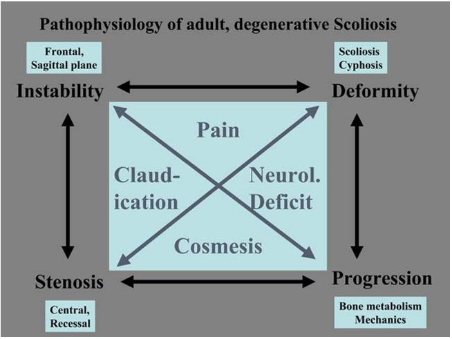
Degenerative adult scoliosis, specifically in the lumbar
spine, is characterized by quite a uniform pathomor-
phology and pathomechanism. The asymmetric degen-
eration of the disc and/or the facet joints leads to an
asymmetric loading of the spinal segment and conse-
quently of a whole spinal area. This again leads to an
asymmetric deformit y, for example, scoliosis and/or
kyphosis. Such a deformity again triggers asymmetric
degeneration and induces asymmetric loading, creating
a vicious circle (Fig. 9) and enhancing curve progres-
sion. On the one hand, the curve progression is given
by the patho mechanism of an adult degenerative curve,
and on the other hand by the specific bone metabolism
of the post-menopause female patients with a certain
degree of osteoporosis, who are most frequently af-
fected by the degenerative form of scoliosis. The po-
tential of individual asymmetric deformation and
collapse in the weak osteoporotic vertebra is clearly
increased and cont ributes further to the curve pro-
gression.
The destruction of discs, facet joint s and joint
capsules usually ends in some form of uni- or multi-
segmental sagittal and/or frontal latent or obvious
instability. There may be not only a spondylolisthesis,
meaning a slip in the sagittal plain, but also transla-
tional dislocations in the frontal plain or rather three-
dimensionally when expressing itself in a rotational
dislocation (Figs. 1, 3, 6, 15). The biological reaction
to an unstable joint or, in the case of the spine, an
unstable segment, is the formation of osteophytes at
the facet joints (spondylarthritis) and at the vertebral
endplates (spondylosis), both contributing to the
increasing narrowing of the spinal canal together with
Fig. 2 Secondary changes in degenerative scoliosis: facet joint
hypertrophy, recessal stenosis
929

Citations
More filters
Journal ArticleDOI

Compensatory mechanisms recruited against proximal junctional kyphosis by patients instrumented from the thoracolumbar junction to the iliac

TL;DR: In this paper, the authors performed a retrospective analysis of data collected prospectively in a multicenter adult spinal deformity database and found that patients suffering from PJK/PJF after (T8-L2) to iliac instrumentation with minimum two-year follow-up had greater capacity to recruit compensatory mechanisms against sagittal imbalance.
Journal ArticleDOI

Three-dimensional alignment changes of the shoulder girdle between the supine and standing positions

TL;DR: It was showed that angular rotations and positions of the clavicle and scapula change significantly with position due to the effect of gravity.
Journal ArticleDOI

Preoperative and postoperative sagittal plane analysis in adult idiopathic scoliosis in patients older than 40 years of age

TL;DR: It is seen that worse sagittal profile leads to worse HRQOL, but after multivariate analysis, only spinal tilt (ST) persisted as possible predictor for worse SRS activity scores.
Journal ArticleDOI

Radiological Evaluation and Classification of Adult Spinal Deformity

TL;DR: In order to maintain spinal balance, the combination of thoracic kyphosis, lumbar lordosis, and pelvic tilt should be harmonized, which is important to maintaining a balanced spine.
Journal ArticleDOI

Was ist eigentlich eine adulte spinale Deformität

TL;DR: The adulte spinale deformitat (ASD) as mentioned in this paper is ein komplexes Krankheitsbild, which is used in Wirbelsaulenchirurgen.
References
More filters
Journal ArticleDOI

The Impact of Positive Sagittal Balance in Adult Spinal Deformity

TL;DR: This study shows that although even mildly positive sagittal balance is somewhat detrimental, severity of symptoms increases in a linear fashion with progressive sagittal imbalance, and shows that kyphosis is more favorable in the upper thoracic region but very poorly tolerated in the lumbar spine.
Journal ArticleDOI

Morbidity and mortality in association with operations on the lumbar spine. The influence of age, diagnosis, and procedure.

TL;DR: Complications, length of hospitalization, and charges were higher for patients who had had a spinal arthrodesis than for those who had not, and operations for conditions other than a herniated disc were associated with more complications and greater use of resources than were operations for removal of a hernia.
Journal ArticleDOI

Morbidity and Mortality in Association with Operations on the Lumbar Spine: The Influence of Age, Diagnosis, and Procedure

TL;DR: Complications, length of hospitalization, and charges were higher for patients who had had a spinal arthrodesis than for those who had not, and operations for conditions other than a herniated disc were associated with more complications and greater use of resources than were operations for removal of a hernia.
Journal ArticleDOI

Natural history of untreated idiopathic scoliosis after skeletal maturity.

TL;DR: In an unselected group of patients with severe curves a mortality rate of 17% was found, twice as much as in the Italian general population, and the cosmetic appearance of these patients at long-term follow-up was better than that at the end of growth, even though the curves progressed.
Related Papers (5)
Frequently Asked Questions (12)
Q1. What are the contributions in this paper?

In this paper, the authors defined scoliosis as a spinal deformity in a skeletally mature patient with a Cobb angle of more than 10 in the coronal plain. 

The non-surgical treatment options [9, 20, 34, 46, 67] consist basically of non-steroid anti-inflammatory medication, muscle relaxants, pain medication, muscle exercises, swimming and occasionally gentle traction, while avoiding manipulations and physical activation that may increase the pain. 

depending on the cause of the curve, the lumbosacral junction usually is degenerated: disc space narrowing, facet joint arthritis, vertebral obliquity and possibly rotational deformity and sometimes even spontaneous fusion of L5 to S1 might be a consequence of a lumbosacral transitional anomaly or a progressed degeneration. 

The asymmetric degeneration of the disc and/or the facet joints leads to an asymmetric loading of the spinal segment and consequently of a whole spinal area. 

The second important symptom of adult degenerative scoliosis is radicular pain and claudication symptoms when standing or walking [57, 73]. 

As patients, who present themselves with significant clinical problems in the context of adult scoliosis, get older, minimal invasive procedures to address exactly the most relevant clinical problem may become more and more important, basically ignoring the overall deformity and degeneration of the spine. 

The posterior pedicular systems nowadays allow a powerful manipulation, correction, and stabilization of the lumbar spine, as long as a proper posterior release precedes the corrective and stabilization procedure. 

Whether the emerging dynamic fixation devices or even disc arthroplasty will be an option in the surgical treatment of adult degenerated scoliosis remains to be considered as more experience is acquired with that kind of implant. 

The sagittal deformity is almost always exclusively a flat back syndrome or a loss of physiological lordosis and in extreme situations a real kyphosis. 

This is the time when degenerative scoliosis may become increasingly symptomatic because the curve may progress due to the asymptomatic load on weakened vertebrae, which get more wedged and deformed. 

When analyzed, regarding their overall daily activity by different questionnaires [50], most of these patients irrespective of age have improved in almost all categories of quality of life, and the use of regular pain medication is reduced substantially in more than 70% of these patients. 

Keywords Adult scoliosis Æ Degenerative scoliosis Æ Spinal stenosis Æ Adult deformity Æ Secondary scoliosistherefore the main bulk of adult scoliosis. 

Trending Questions (1)
What are the physical causes of adult degenerative scoliosis?

Physical causes of adult degenerative scoliosis include asymmetric degeneration leading to increased load, facet joint destruction, spinal stenosis, and osteoporosis, especially in post-menopausal females.