
All figures (9)
 Figure 3. Spongistatin 1 inhibits migration of HUVECs. A) Spongistatin 1, vinblastine, CA4P, and paclitaxel inhibited wound closure of a scratched HUVEC monolayer in a dose-dependent manner. *P 0.05; 1-way ANOVA. B) Representative images of quantification of wound healing. Cell-free area is gray. C) Actin cytoskeleton in migrating HUVECs: under spongistatin 1 treament, formation of lammelipodia is reduced, and formation of cortical rings is increased.
 Figure 2. Spongistatin 1 exerts strong antiproliferative effects on HUVECs in nontoxic concentrations. A) Cytotoxic concentrations of spongistatin 1 were determined by metabolic conversion of resazurin to resorufin in confluent and proliferative HUVECs. Significantly reduced metabolic activity could be observed at a concentration of spongistatin 1 as low as 5.0 nM in proliferative cells, whereas in confluent HUVECs, a significant reduction could be determined at 50.0 nM of spongistatin 1. B–D) Effects of spongistatin 1 on apoptosis(B), proliferation (C), and cell cycle (D) were analyzed in proliferating HUVECs in comparison with established tubulin antagonists vinblastine, CA4P, and paclitaxel. *P 0.05; 1-way ANOVA.
 Figure 4. Spongistatin 1 reduces directional migration in HUVECs. A) Track plots of control cells and spongistatin 1-treated cells of representative chemotaxis experiments. Starting point of each single cell is placed in the center of the diagram. Tracks of cells moving toward FCS are black;
 Figure 8. Spongistatin 1 and nocodazole disrupt translocation of PKC . A) Detection of the PKCisoforms , , and ε in membranous and cytoplasmic fractions of HUVECs that were either stimulated with 10 nM PMA for 2 h (PMA), or preincubated with 5 nM spongistatin 1 before PMA-stimulus (SP1). Detection of VE-cadherin (VEC) and -actin demonstrated purity of the fractions and equal protein loading. B) To investigate whether inhibition of translocation of PKC is a microtubule-dependent effect, experiment was repeated with established microtubule-disrupting agent nocodazole (preincubation with 10 M before PMA stimulus). *P 0.05 vs. PMA; rank-sum test.
 Figure 5. Tube formation and endothelial sprouting are inhibited by spongistatin 1. A) Representative images of tube formation. S.CORE tube formation analysis determines cellular structures as tubes (dark gray) and nodes (light gray). B) Tube formation of endothelial cells on Matrigel was reduced to half by 1.0 nM spongistatin 1 (SP1), 5.0 nM vinblastine (vinb), 10.0 nM CA4P, and 50.0 nM paclitaxel (pacl). *P 0.05; 1-way ANOVA. C) Representative pictures of aortic ring assay showing vascular sprouting of the 3 treatment groups (control and 0.5 and 1.0 nM spongistatin 1).
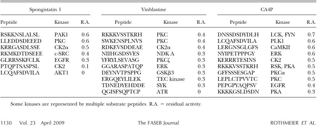 TABLE 1. Kinome array data showing kinases that were reduced most in their activity in tubulin-antagonist-treated HUVECs compared to control cells
 Figure 6. Spongistatin 1 inhibits neovascularization of mouse corneas. A) Corneal neovascularization was examined and photographed on day 6 after implantation of pellets containing bFGF ( 20 view of mouse eye; dots encircle area of implanted pellet). Corneal neovascularization in spongistatin 1-treated animals was markedly reduced compared to vehicletreated control animals (control). B, C) Angiogenic responses were measured as clock hours (B) and vessel length of neovascularization (C). *P 0.001; Student’s t test.
 Figure 7. Spongistatin 1 inhibits phosphorylation of PKC substrates and translocation of PKC without directly blocking PKC activity. A) Western blot analysis to investigate phosphorylation activity of PKC in spongistatin 1-treated cells (same stimulation as in PepChip), using an antibody against phosphorylated PKC substrates (serine residues). Kinase inhibitor staurosporine (stauro) abolished phosphorylation of PKC substrates. In contrast, spongistatin 1 reduced phosphorylation only of distinct PKC substrates (arrows). B) In PKC in vitro assay, spongistatin 1 did not significantly inhibit phosphorylation activity of PKC isoforms , I, II, , ε, or (P 0.05; rank-sum test).
 Figure 1. Live-cell imaging and tubulin fractionation proved the disassembly of microtubules by spongistatin 1. A) GFP-tubulin-transfected HUVECs were stimulated with 2.0 nM spongistatin 1 for 16 h. Bottom panels show magnification of microtubules of the HUVECs. B) Cell lysates of paclitaxel (pacl)- and spongistatin 1 (SP1)-stimulated HUVECs were separated in PIPES buffer-soluble and -insoluble fractions. Tubulin heterodimers were detected in the soluble fraction, polymerized microtubules in the insoluble fraction.


