scispace - formally typeset
Open AccessPosted ContentDOI

Characterization of SARS2 Nsp15 Nuclease Activity Reveals it's Mad About U

TLDR
In this article, the authors used cryo-EM to capture structures of Nsp15 bound to RNA in pre-and post-cleavage states, and determined how the sequence of the RNA substrate dictates cleavage and found that outside of polyU tracts, Nsp 15 has a strong preference for purines 3 of the cleaved uridine.
Abstract
Nsp15 is a uridine specific endoribonuclease that coronaviruses employ to cleave viral RNA and evade host immune defense systems. Previous structures of Nsp15 from across Coronaviridae revealed that Nsp15 assembles into a homo-hexamer and has a conserved active site similar to RNase A. Beyond a preference for cleaving RNA 3 of uridines, it is unknown if Nsp15 has any additional substrate preferences. Here we used cryo-EM to capture structures of Nsp15 bound to RNA in pre- and post-cleavage states. The structures along with molecular dynamics and biochemical assays revealed critical residues involved in substrate specificity, nuclease activity, and oligomerization. Moreover, we determined how the sequence of the RNA substrate dictates cleavage and found that outside of polyU tracts, Nsp15 has a strong preference for purines 3 of the cleaved uridine. This work advances our understanding of how Nsp15 recognizes and processes viral RNA and will aid in the development of new anti-viral therapeutics.

read more

Content maybe subject to copyright    Report

Characterization of SARS2 Nsp15 Nuclease Activity Reveals
it’s Mad About U
Meredith N. Frazier
1
, Lucas B. Dillard
2
, Juno M. Krahn
2
, Lalith Perera
2
, Jason G. Williams
3
, Isha M.
Wilson
1
, Zachary D. Stewart
1
, Monica C. Pillon
1
, Leesa J. Deterding
3
, Mario J. Borgnia
2
, Robin E.
Stanley
1*
1
Signal Transduction Laboratory, National Institute of Environmental Health Sciences, National
Institutes of Health, Department of Health and Human Services, 111 T. W. Alexander Drive, Research
Triangle Park, NC 27709, USA
2
Genome Integrity and Structural Biology Laboratory, National Institute of Environmental Health
Sciences, National Institutes of Health, Department of Health and Human Services, 111 T. W.
Alexander Drive, Research Triangle Park, NC 27709, USA
3
Epigenetics and Stem Cell Biology Laboratory, National Institute of Environmental Health Sciences,
National Institutes of Health, Department of Health and Human Services, 111 T. W. Alexander Drive,
Research Triangle Park, NC 27709, USA
*To whom correspondence should be addressed. Tel: 01 984 287 3568; Email: robin.stanley@nih.gov
Present Address: Monica C. Pillon, Department of Biochemistry and Molecular Biology, Baylor
College of Medicine, Houston, TX, 77030, USA
ABSTRACT
Nsp15 is a uridine specific endoribonuclease that coronaviruses employ to cleave viral RNA and
evade host immune defense systems. Previous structures of Nsp15 from across Coronaviridae
revealed that Nsp15 assembles into a homo-hexamer and has a conserved active site similar to
RNase A. Beyond a preference for cleaving RNA 3′ of uridines, it is unknown if Nsp15 has any
additional substrate preferences. Here we used cryo-EM to capture structures of Nsp15 bound to
RNA in pre- and post-cleavage states. The structures along with molecular dynamics and biochemical
assays revealed critical residues involved in substrate specificity, nuclease activity, and
oligomerization. Moreover, we determined how the sequence of the RNA substrate dictates cleavage
and found that outside of polyU tracts, Nsp15 has a strong preference for purines 3′ of the cleaved
uridine. This work advances our understanding of how Nsp15 recognizes and processes viral RNA
and will aid in the development of new anti-viral therapeutics.
USC 105 and is also made available for use under a CC0 license.
(which was not certified by peer review) is the author/funder. This article is a US Government work. It is not subject to copyright under 17
The copyright holder for this preprintthis version posted June 1, 2021. ; https://doi.org/10.1101/2021.06.01.446181doi: bioRxiv preprint

INTRODUCTION
The novel SARS-CoV-2 (severe acute respiratory syndrome coronavirus 2) emerged in late 2019 and
became a worldwide pandemic that is still ongoing and has infected millions worldwide (1).
Coronaviruses are members of the Nidovirus order, which encompasses large, positive-strand RNA
viruses with genomes that range in size from 12-41 kb (2). The 30 kb SARS-CoV-2 genome encodes
for 4 structural proteins that are part of the mature viral particle, 8 accessory proteins, and 15 non-
structural proteins (Nsps) (3). The Nsps are encoded in two open reading frames found in the first
two-thirds of the viral genome. These proteins are translated by host ribosomes as two long
polyproteins and are cleaved into functional proteins by the viral proteases (4). The Nsps play
important roles in viral replication and pathogenicity and many of them are promising drug targets
(3,4).
Nsp15 is a uridine specific endoribonuclease conserved across the Coronaviridae family (5).
Enzymatic activity occurs in the C-terminal EndoU domain, which is more broadly conserved across
nidoviruses, suggesting that this endoribonuclease activity is critically important for large, positive-
strand RNA viruses (5,6). Work in animals and cell culture has shown that Nsp15 function is not
necessary for viral replication, however Nsp15 nuclease activity is critically important for evasion of
the host immune response to the virus, specifically by preventing the activation of dsRNA sensors (7-
11). For example, in studies of porcine endemic diarrhea virus (PEDV), Nsp15-deficient virus resulted
in higher levels of type I and III interferon responses in cells, and piglets infected with the mutant virus
had much higher survival rates than those infected with WT PEDV (8). A similar effect was also seen
in studies of mouse hepatitis virus (MHV); mice immunized with Nsp15 nuclease deficient virus were
able to successfully clear WT virus, with commonly affected organs showing no pathology (7). Recent
work also revealed a similar trend in the chicken infection bronchitis virus (IBV), where animals
infected with nuclease-deficient virus had reduced mortality and viral shedding (12). Therefore, Nsp15
is a promising therapeutic target for coronaviruses.
One of the major outstanding questions about the function of Nsp15 is what is its RNA target for
cleavage (5). Recent studies have begun to shed light on this important question (9,13). One study
found a link between Nsp15 activity and the length of the polyuridine sequence at the 5¢ end of the
template negative strand. When Nsp15-mutant MHV infected cells, there was a greater amount of
polyuridine (PUN) RNA compared to cells infected with WT virus, suggesting Nsp15 cleaves the PUN
RNA produced in the negative strand intermediate state (9). The PUN RNA was found to trigger the
pathogen associated molecular pattern (PAMP) receptor MDA5, which mediates interferon response
(9). Another study used cyclic phosphate RNA sequencing to identify Nsp15 cleavage products within
MHV infected bone marrow-derived macrophages (13). This analysis revealed that Nsp15 cleaves
numerous targets throughout the positive strand with a preference for cleaving between U^A and C^A
sequences (13). More recent work with IBV demonstrated that Nsp15 nuclease activity prevents the
accumulation of both dsRNA and cytoplasmic stress granules which have established anti-viral
USC 105 and is also made available for use under a CC0 license.
(which was not certified by peer review) is the author/funder. This article is a US Government work. It is not subject to copyright under 17
The copyright holder for this preprintthis version posted June 1, 2021. ; https://doi.org/10.1101/2021.06.01.446181doi: bioRxiv preprint

properties (14). Collectively these studies confirm that Nsp15 nuclease activity is critical to prevent the
accumulation of viral ds-RNA and activation of the immune response.
Numerous structures of Nsp15 have been determined from several Coronaviridae family members,
however there are no structures of Nsp15 with more than a di-nucleotide bound, which has hindered
our understanding of how Nsp15 recognizes its RNA targets. Crystal structures of Nsp15 revealed
that Nsp15 assembles into a hexameric complex, formed from back-to-back trimers with the EndoU
domains facing outward (15-19). The active site of Nsp15 shares considerable similarity to the well-
studied endoribonuclease RNase A, and is composed of a catalytic triad including two histidines and
lysine (18,20,21) . These residues support a two-step reaction of transesterification and hydrolysis,
however due to an altered position of one active site histidine, Nsp15 accumulates products from the
transesterification reaction which contain a cyclic phosphate (20). Recent structures determined of
Nsp15 bound to uridine nucleotides uncovered the molecular basis for uridine specificity, which is
driven by a well-conserved serine residue within the uridine binding pocket (20,21). However, beyond
the preference for uridines, it is unclear if Nsp15 has any additional specificity requirements. RNA
sequencing suggests there is a preference for adenine 3¢ to the uridine, but the structural basis for
this is unknown (13). In contrast, RNase A is known to have additional non-catalytic sites that affect
substrate preference (22,23), prompting further characterization of how Nsp15 engages RNA.
Here we used cryo-EM, molecular dynamics simulations, and in vitro RNA cleavage assays to probe
the substrate specificity of SARS-CoV-2 Nsp15. We determined cryo-EM reconstructions of Nsp15
with RNA bound in the pre- and post-cleavage states. The structures revealed that, in contrast to
RNase A, Nsp15 does not contain any additional well-ordered sites for RNA binding and recognition.
This observation was further supported by molecular dynamics simulations with tri-nucleotide
substrates. We probed RNA specificity by determining how the nucleotide 5¢ and 3¢ of the uridine
affects cleavage and found that Nsp15 has a preference for purines 3′ of the cleaved pyrimidine.
Finally, we looked at Nsp15’s ability to cleave SARS-CoV-2 viral RNA substrates, such as the PUN
and the transcriptional regulatory sequence (TRS). Collectively our work suggests that SARS CoV-2
Nsp15 is able to cleave a broad spectrum of RNA substrates and that this activity is driven by
recognition of uridine within the active site.
MATERIAL AND METHODS
Protein expression and purification
Wild type (WT) and mutant Nsp15 constructs were created as described previously (20). Nsp15 was
overexpressed in E. coli C41 (DE3) competent cells in Terrific Broth with 100 mg/L ampicillin. At an
optical density (600 nm) between 0.8-1.0, cultures were cooled at 4˚C for 1 hour prior to induction with
0.2 mM Isopropyl β-D-1-thiogalactopyranoside (IPTG). Cells were harvested after overnight
expression at 16˚C and stored at -80˚C until use. Nsp15 purification was done as described
previously (20). Briefly, cells were resuspended in Lysis Buffer (50 mM Tris pH 8.0, 500 mM NaCl, 5%
glycerol, 5 mM b-ME, 5 mM imidazole) supplemented with cOmplete EDTA-free protease inhibitor
USC 105 and is also made available for use under a CC0 license.
(which was not certified by peer review) is the author/funder. This article is a US Government work. It is not subject to copyright under 17
The copyright holder for this preprintthis version posted June 1, 2021. ; https://doi.org/10.1101/2021.06.01.446181doi: bioRxiv preprint

tablets (Roche) and disrupted by sonication. The lysate was clarified at 26,915 x g for 50 minutes at
4˚C and then incubated with TALON metal affinity resin (Clontech). His-Nsp15 was eluted from the
resin with 250 mM imidazole, and buffer exchanged into Thrombin Cleavage Buffer (50 mM Tris pH
8.0, 150 mM NaCl, 5% glycerol, 2 mM b-ME, 2 mM CaCl
2
) for cleavage at room temperature for 3
hours. The cleavage reaction was repassed over TALON resin and quenched with 1 mM
phenylmethylsulfonyl fluoride (PMSF) prior to gel filtration using a Superdex-200 column equilibrated
in SEC buffer (20 mM Hepes pH 7.5, 150 mM NaCl, 5 mM MnCl
2
, 5 mM b-ME).
Cryo-EM sample preparation
Purified Nsp15 was diluted in a low-salt buffer (20 mM Hepes pH 7.5, 100 mM NaCl, 5 mM MnCl
2
, 5
mM b-ME) to 0.75 µM and incubated with excess RNA substrates (1 mM AU
f
A or AUA, see Table S1)
for 1 hour at 4˚C. UltrAuFoil R1.2/1.3 300 mesh gold grids (Quantifoil) were plasma cleaned (Pie
Scientific) before use. The Nsp15/RNA mixture (3 µL) was deposited onto the grids, back-blotted for 3
seconds, and vitrified using an Automatic Plunge Freezer (Leica).
Data collection and processing
Nsp15 images were collected using a Krios electron microscope at 300 keV with a Gatan K2 detector
in super-resolution mode. Beam-induced motion and drift were corrected using MotionCor2 (24) and
aligned dose-weighted images were used to calculate CTF parameters using CTFFIND4 (25).
CryoSPARC v2 (26) was used in all subsequent image processing. Particles were selected by
template-based particle picking, downsampled by a factor of 4, extracted with a box size of 64 and
subjected to an initial round of 2D classification. Full resolution particle projections from good classes
were re-extracted using a box size of 256. Ab initio reconstruction was used to generate an initial
model. Three independent 3D refinement cycles were performed while applying C1, C3, and D3
symmetry respectively. Although previous apo and UTP-bound datasets had D3 symmetry, the longer
RNA bound in both datasets here resulted in particles that no longer had D3 symmetry, perhaps due
to incomplete or mixed occupancy. Inspection of the C1 map did not reveal any asymmetric
differences, although active site density was difficult to interpret for one half of the pre-cleavage state
map. Therefore, C3 symmetry was used for model building and analysis for both datasets. Maps were
re-scaled to optimize RMS fit to core domain residues of reference structure PDBID 6WLC (21).
Model building
A SARS-CoV-2 Nsp15 crystal structure (PDBID 6WLC) was used as a starting model and fit into the
cryo-EM maps using rigid body docking in Phenix (27). For the pre-cleavage state, which was
captured with an AU
f
A tri-nucleotide, the density for the 5¢ A was weaker than the density for the U, so
only the C5¢ group was modeled; no density was observed for the 3¢ A. For the post-cleavage state,
the 5¢ A could be fit in the density along with the U. A combination of rigid body and real-space
refinement in Phenix as well as iterative rounds of building in COOT (28) were used to improve the fit
of the model. Molprobity (29) was used to evaluate the model (Table 1). Figures were prepared using
Chimera (30) and Chimera X (31).
USC 105 and is also made available for use under a CC0 license.
(which was not certified by peer review) is the author/funder. This article is a US Government work. It is not subject to copyright under 17
The copyright holder for this preprintthis version posted June 1, 2021. ; https://doi.org/10.1101/2021.06.01.446181doi: bioRxiv preprint

FRET endoribonuclease assay
Nsp15 cleavage was monitored in real-time as described previously (19,20). Briefly, 6-mer substrates
were labeled with 5′-fluorescein (FI) and 3′-TAMRA, where TAMRA quenches FI and cleavage is
measured by increasing FI fluorescence (5′-FI-AAxxxA-TAMRA-3′; x nucleotides varied among
substrates) (see Supplementary Table 1). The substrate (0.8 µM) was incubated with Nsp15 (2.5 nM)
in RNA cleavage buffer (20 mM Hepes pH 7.5, 75 mM NaCl, 5 mM MnCl
2
, 5 mM DTT) at 25°C for 60
minutes. Fluorescence was measured every 2.5 minutes using a POLARstar Omega plate reader
(BMG Labtech) set to excitation and emission wavelengths of 485 ± 12 nm and 520 nm, respectively.
Three technical replicates were performed for each condition, and the assay was repeated with at
least two independent protein preparations. Prism (Graphpad) was used to calculate significant
differences using Dunnett’s T3 multiple corrections test.
Urea-PAGE endoribonuclease assay
Double fluorescently-labeled RNA substrates (5′-FI and 3′-Cy5, 500 nM) were incubated with Nsp15
(50 nM) in RNA cleavage buffer (20 mM Hepes pH 7.5, 150 mM NaCl, 5 mM MnCl
2
, 5 mM DTT, 1
u/µL RNasin ribonuclease inhibitor) at room temperature for 30 minutes, with samples collected at 0,
1, 5, 10, and 30 minutes. The reaction was quenched with 2x urea loading buffer (8M urea, 20 mM
Tris pH 8.0, 1 mM EDTA). Due to the expected size of cleavage products and the size of
bromophenol blue, loading buffer without dye was used. To monitor the gel front, a control lane of
protein only with bromophenol blue was run. To generate a ladder, alkaline hydrolysis of the RNA was
carried out for 15 min at 90˚C using 1 µM RNA in alkaline hydrolysis buffer (50 mM sodium carbonate
pH 9.2, 1 mM EDTA) and quenched with 2x urea loading buffer. The cleavage reactions were
separated using 15%-20% TBE-urea PAGE gels and visualized with a Typhoon RGB imager
(Amersham) using Cy2
ex
=488 nm, λ
em
=515-535 nm
) and Cy5
ex
=635 nm, λ
em
=655-685 nm)
channels.
Mass spectrometry of RNA cleavage products
Mass spectrometry was performed as previously described (20). Briefly, the FRET RNA substrate of
interest (0.8 µM) was incubated +/- Nsp15 (2.5 nM) in RNA cleavage buffer for 30 minutes at RT. For
mass spectrometry analysis, the reaction was chromatographically separated with a gradient of buffer
A (400 mM hexafluoro-2-propanol, 3 mM triethylamine, pH 7.0) and buffer B (methanol). Parallel
reaction monitoring (PRM) analyses were included in the MS analyses with included masses of m/z
914.14; 923.14; 1463.42.
Molecular dynamics simulations
Based on the RNA bound cryo-EM hexamer structure of Nsp15, the initial structure of Nsp15-AUA
hexamer complex was prepared by manually introducing an adenine nucleotide at the B
-2
position.
Except for H250, all histidine residues were selected to be N
ε
protonated. Since the ring nitrogen
atoms on H250 were found to make two strong hydrogen bonds with the phosphate backbone and the
USC 105 and is also made available for use under a CC0 license.
(which was not certified by peer review) is the author/funder. This article is a US Government work. It is not subject to copyright under 17
The copyright holder for this preprintthis version posted June 1, 2021. ; https://doi.org/10.1101/2021.06.01.446181doi: bioRxiv preprint

Figures
Citations
More filters
Journal ArticleDOI

Mechanisms of Antiviral Immune Evasion of SARS-CoV-2.

TL;DR: In this paper, a detailed understanding of the host-pathogen interactions that unbalance the IFN response is critical for the design and deployment of host-targeted therapeutics to manage COVID-19.
Journal ArticleDOI

Mechanisms of Antiviral Immune Evasion of SARS-CoV-2

TL;DR: In this article , a detailed understanding of the host-pathogen interactions that unbalance the IFN response is critical for the design and deployment of host-targeted therapeutics to manage COVID-19.
References
More filters
Journal ArticleDOI

SARS-Coronavirus Replication Is Supported by a Reticulovesicular Network of Modified Endoplasmic Reticulum

TL;DR: The ultrastructural description of this “replication network” will aid to further dissect the early stages of the coronavirus life cycle and its virus-host interactions.
Journal ArticleDOI

RNA Replication of Mouse Hepatitis Virus Takes Place at Double-Membrane Vesicles

TL;DR: The ultrastructural analysis revealed double-membrane vesicles (DMVs) in the cytoplasm of mouse hepatitis virus-infected cells, concluding that the DMVs carry the MHV RNA replication complex and are the site ofMHV RNA synthesis.
Journal ArticleDOI

Emerging Pandemic Diseases: How We Got to COVID-19.

TL;DR: This work states that disease emergence reflects dynamic balances and imbalances, within complex globally-distributed ecosystems comprised of humans, animals, pathogens, and the environment, which is a necessary step in controlling future devastating disease emergences.
Journal ArticleDOI

The Proteins of Severe Acute Respiratory Syndrome Coronavirus-2 (SARS CoV-2 or n-COV19), the Cause of COVID-19.

TL;DR: The proteins found in the SARS CoV-2 that caused this global pandemic are reviewed to provide a better understanding of the disease.
Journal ArticleDOI

The molecular virology of Coronaviruses

TL;DR: The molecular virology of coronavirus infection, including its entry into cells, its remarkably sophisticated gene expression and replication mechanisms, its extensive remodeling of the intracellular environment, and its multifaceted immune evasion strategies are presented.
Related Papers (5)
Frequently Asked Questions (14)
Q1. What was used to improve the fit of the model?

A combination of rigid body and real-space refinement in Phenix as well as iterative rounds of building in COOT (28) were used to improve the fit of the model. 

This work advances their understanding of how Nsp15 recognizes and processes viral RNA and will aid in the development of new anti-viral therapeutics. ( which was not certified by peer review ) is the author/funder. This article is a US Government work. 

Since the trinucleotide was not bound to the binding site residues during the entire half a microsecond production runs in most systems, the energy calculations were performed for each 50 ns segments (with 50 samples selected at each nanosecond) separately for each trajectory. 

other viral proteins may influence Nsp15 RNA targets and regulation in host cells, as Nsp15 is believed to localize within the replication-transcription complex of Nsps, including the RdRp complex (54,55). 

Enzymatic activity occurs in the C-terminal EndoU domain, which is more broadly conserved across nidoviruses, suggesting that this endoribonuclease activity is critically important for large, positivestrand RNA viruses (5,6). 

Nsp15 is a key player in blocking activation of host dsRNA sensors by preventing the accumulation of viral RNA and a promising therapeutic target (9,13,14). 

Work in animals and cell culture has shown that Nsp15 function is not necessary for viral replication, however Nsp15 nuclease activity is critically important for evasion of the host immune response to the virus, specifically by preventing the activation of dsRNA sensors (7- 11). 

Rootmean square deviations (RMSDs) were used to establish the stability of the simulated systems (Supplementary Figure 4A) in which the isolated monomer systems displayed elevated dynamics (as assessed by RMSDs) compared to the protomers assembled into the hexamer. 

The authors captured the post-cleavage state by incubating Nsp15 with excess unmodified RNA (AUA) prior to vitrification and cryo-EM data collection. 

The authors incubated WT Nsp15 with the TRS-N and TRS-S containing RNA substrates and then resolved the cleavage products on denaturing urea gels. 

Based on these endoribonuclease assay results, the authors define the consensus motif for Nsp15 cleavage as (N)(U)^(R>U>>C) (where N is any nucleotide and R is a purine). 

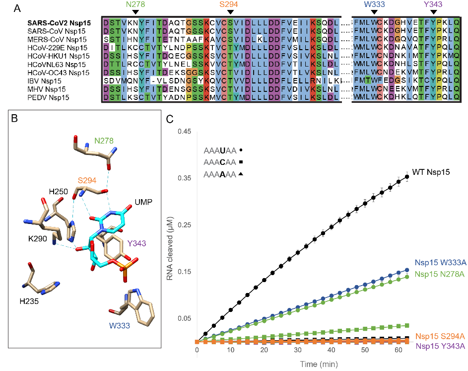
Consistent with their assay results with a six nucleotide substrate, the N278A Nsp15 variant cleaved more slowly and produced more C cleavage products than WT Nsp15 (Fig. 7). 

the authors also looked at Nsp15’s ability to degrade polyU sequences and found that Nsp15 efficiently degrades polyU containing RNAs in vitro. 

the authors turned to molecular dynamics simulations to further characterize the behavior of the nucleotides near the active site.